Uworld Notes
Basic Science
Reticulocytes
_..
The reticulocyte is an immature RBC that is slightly larger and bluer than a mature RBC. It lacks a cell nucleus but retains a basophilic, reticular (mesh-like) network of residual ribosomal RNA. The ribosomal RNA appears blue microscopically after the application of the Wright-Giemsa stain. After spending a day or so in the bloodstream, the reticulocytes are transformed into mature red blood cells that have a lifespan of approximately 120 days.
Collagen
_..

Evolution of a myocardial infarction occurs in several stages. The final stage of the healing process begins 2 weeks after infarction and involves increased type I collagen deposition. Fibrosis continues until about 2 months after infarction, resulting in a dense collagenous scar composed primarily of type I collagen.
Anatomy
Thoracocentisis
_..

Biochemistry
Vitamin A
_..
Vitamin A deficiency result:
goblet/columnar epithelium of conjunctiva metaplasia into keratinizing squamous.
night blindness (immune deficiency)
APML 15:17 (immune maturation)
O2 radical
_..
O2 to O2- to H2O2 to OH- to H2O
O2- converted by superoxide dismutase to H2O2
H2O2 converted to water and O2 by catalase
OH- converted by glutathione to water
Collagen and vitamins
_..
vit C: hydroxylation of proline/lysine
copper: lysyl oxidase: lysine/hydroxylysine cross linking
zinc: collagenase cofactor, replace type 3 with type 1
Pathology
Scar
_..
hypertrophic scar: excess production of scar tissue, localized to wound. Type 1 collagen
keloid: excess production of scar out of proportion. Type 2 collagen. Risks: AA, earlobe, face
CCL4
organic solvent used in dry cleaning
free radical resulting in cell injury, impaired protein synthesis in liver
decreased apolipoprotein, fatty liver.,
Conus Medularis
_..
In an adult, the spinal cord terminates in a tapering fashion at the conus medullaris at approximately the L2 vertebral level. After this point, spinal nerves from the conus medullaris exit inferiorly through their respective intervertebral foramina. This collection of spinal nerves (now considered peripheral nerves) is referred to as the cauda equina (i.e. horse's tail).
Conus medullaris syndrome
Refers to lesions at L2. It has symptoms of flaccid paralysis of the bladder and rectum, impotence, and saddle (S3-S5 roots) anesthesia. There is usually mild weakness of the leg muscle if the lesion spares both the lumbar cord and the adjacent spinal and lumbar nerve roots. Common causes include disk herniation, tumors, and spinal fractures.
cauda equina syndrome
Typically results from a massive rupture of an intervertebral disk that is capable of causing compression of two or more of the 18 spinal nerve roots of the cauda equina. However, it can also occur due to any trauma or space-occupying lesion of the lower vertebral column. The cauda equina nerve roots provide the sensory and motor innervation of most of the lower extremities, the pelvic floor, and the sphincters.
Classic symptoms of cauda equina syndrome include low back pain radiating to one or both legs, saddle anesthesia, loss of anocutaneous reflex (as in this patient), bowel and bladder dysfunction (S3-S5 roots), and loss of ankle-jerk reflex with plantar flexion weakness of the feet. Of the potentially involved spinal nerves, a lesion involving S2 through S4 will cause the symptoms described in this patient, indicating impairment of the pudendal nerve that innervates the perineum.
Polymyositis
_..
"Endomysial" inflammatory infiltration is characteristic of polymyositis (an inflammatory disease of unknown etiology that affects skeletal muscles). Manifestations are bilateral, symmetric weakness of proximal muscles. Reflexes are normal, as polymyositis involves the muscles, rather than nerves.
Perfusion Injury
_..
Ischemia is characterized by the reduction of blood flow, usually as a result of mechanical obstruction within the arterial system (eg, thrombus). If the flow of blood to the ischemic tissue is restored in a timely manner, those cells that were reversibly injured will typically recover. Sometimes, however, the cells within the damaged tissue will paradoxically die at an accelerated pace through apoptosis or necrosis after resumption of blood flow. This process is termed reperfusion injury, and is thought to occur secondary to one or more of the following mechanisms: 1) oxygen free radical generation by parenchymal cells, endothelial cells, and leukocytes; 2) severe, irreversible mitochondrial damage described as "mitochondrial permeability transition"; 3) inflammation, which attracts circulating neutrophils that cause additional injury; and 4) activation of the complement pathway, causing cell injury and further inflammation. When the cells within heart, brain, or skeletal muscle are injured, the enzyme creatine kinase leaks across the damaged cell membrane and into circulation (as seen in this patient).
ATN
_..

Neonatal Vit K Deficient
_..

intracranial hemorrhage
increased ICP: big head
Metabolic Alkalosis
_..

Reactive vs Malignant LAD
Lymphadenopathy can represent inflammatory changes within the lymph node (reactive hyperplasia) or malignant transformation. Reactive hyperplasia is a broad term that encompasses all benign, reversible enlargement of the lymphoid tissue secondary to an antigenic stimulus. The nodal response to antigenic stimuli is highly variable and can be classified as follicular hyperplasia, sinus hyperplasia, or diffuse hyperplasia. Follicular hyperplasia occurs when the follicles increase in size and number, whereas sinus hyperplasia occurs when the sinuses enlarge and fill with histiocytes. Diffuse hyperplasia is observed when the nodal architecture is diffusely effaced by sheets of lymphocytes, immunoblasts, and macrophages. When malignant transformation occurs (as in lymphomas), the normal lymph node architecture is distorted or effaced by the proliferation of malignant lymphoid cells, often to a greater extent than that seen with reactive hyperplasia. Malignancy-associated effacement of nodal architecture may be follicular or diffuse.
Reactive lymphoid hyperplasia is polyclonal in that it consists of a proliferation of many different cell types within the lymph node. For each type of lymphocyte responding to an antigenic stimulus, multiple genetically-distinct cells of that variety undergo limited monoclonal expansion, leading to an overall polyclonal response. Malignant transformation, in contrast, is monoclonal in that it results from the unchecked proliferation of a single genetically unique cell from only one cell line.
Evaluation for monoclonality of the lymphocyte population is important when lymphoma is suspected. The clonality of a T-cell population is assessed by molecular methods, such as PCR, that examine the rearrangement of T-cell receptor (TCR) genes. If a single allele for the V region of the T-cell receptor predominates in a lymphocytic population, monoclonal proliferation is suspected. The same principle applies when assessing B-cell clonality. Monoclonal rearrangement of the genes for immunoglobulin variable regions is suggestive of a B-cell lymphoma. Of the choices given, monoclonal TCR gene rearrangement is most indicative of malignant processes, especially in the context of appropriate clinical features (i.e., weight loss, night sweats, fever, anorexia).
Cancer
Routes
_..
carcinoma: lymph nodes
sarcoma: hematogenous spread. Some carcinoma
rcc, hepatocellular, follicular of thyroid, choriocarcinoma
ovarian: body cavity, omentum cake
_..

_..
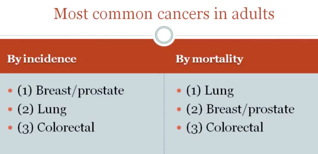
exclude skin (squamous, basal cell)
Carcinogen
_..

alkylating: secondary leukemia/lymphoma
alcohol/tobac co: oropharyngeal carcinoma
alcohol: chronic pancreatitis and hepatitis: pancreatic/hepatocellular carcinoma
arsenic: women apply arsenic to skin to lighten skin, squamous cell carcinoma. Arsenic poisoning in fingernails/hair
arsenic: in cigarette smoke, lung cancer
urothelium of kidney/bladder: cigarette carcinogen concentrate in urine in bladder/kidney
Virus and Radiation
_..

EBV: nasopharyngeal carcinoma, Chinese male, individual in africa; neck mass
Kaposi: endothelium tumor. Older eastern european males (excise), AIDS patients (treat virus, boost T), transplants (reduce immunosuppression)
children: papillary carcinoma of thyroid
nonionizing: nicks in pyrimidine dimers
Protooncogenes
_..

overexpression of PDGFB (over division of cells) drives astrocytoma expression. Astrocytes make more PDGFB
increased receptor: stimulus result in large amount of signal transduction
RET: medullary carcinoma of thyroid association with MEN 2a/2b (prophylactic thyrectomy if positive)
KIT: GIST
Signal transducers: messenger mutated and keep sending signal to nucleus
RAS: commonly mutated in large amount of cancers. If GAP (cleaves GTP, phosphate) is mutated, RAS can't shut off
Nuclear Regulators: TF go to nucleus, upregulate genes
burkitt: cancer of B cells. B cell has IgH (heavy chain) on Ch 14. Translocation causes myc to be massively overproduced. Cells in burkitt grow so rapidly, dying, and eaten up by macrophages (starry sky)
IgH on Ch 14 (B cell cancers)

Benign vs malignant
_..
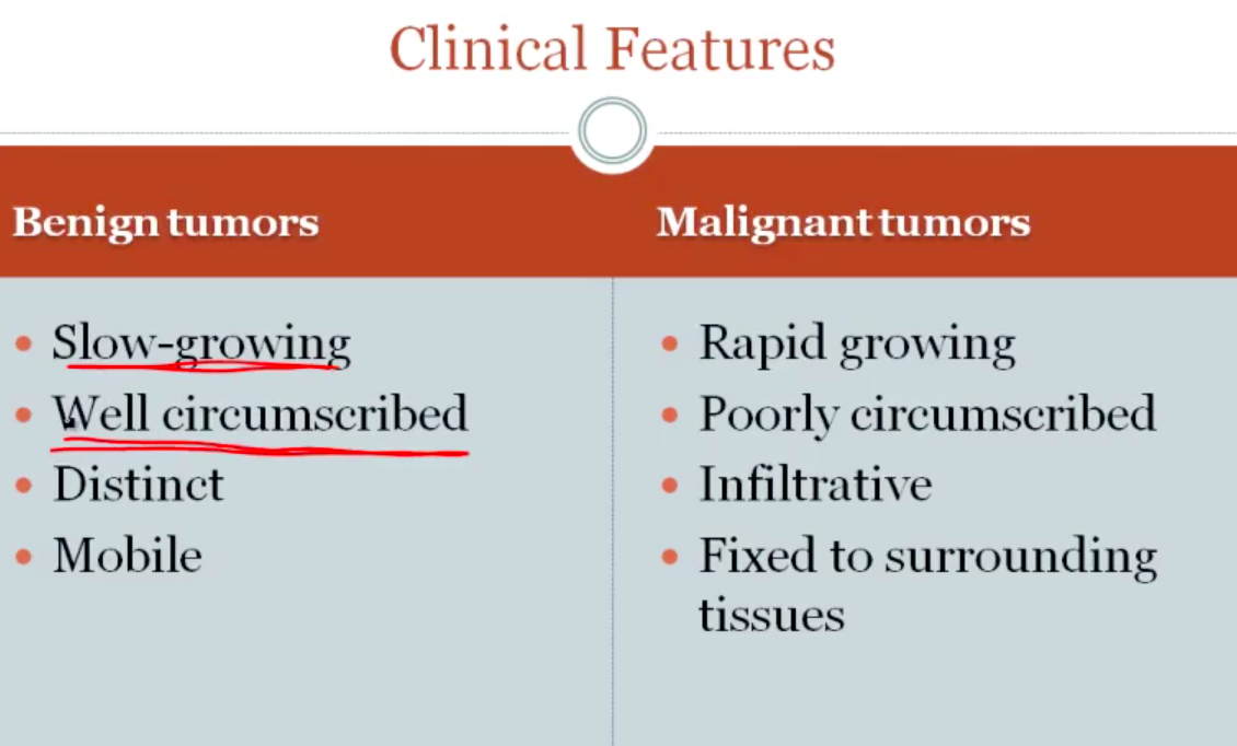

benign: look very similar to surrounding tissues, small nucleus
nuclear pleomorphism: some nucleus big, some small, dark blue
malignant: most cell comprised of nucleus: nucleus rapidly transcribing

left; normal. All cells same size, nucleus
right: anaplastic
Stains
_..
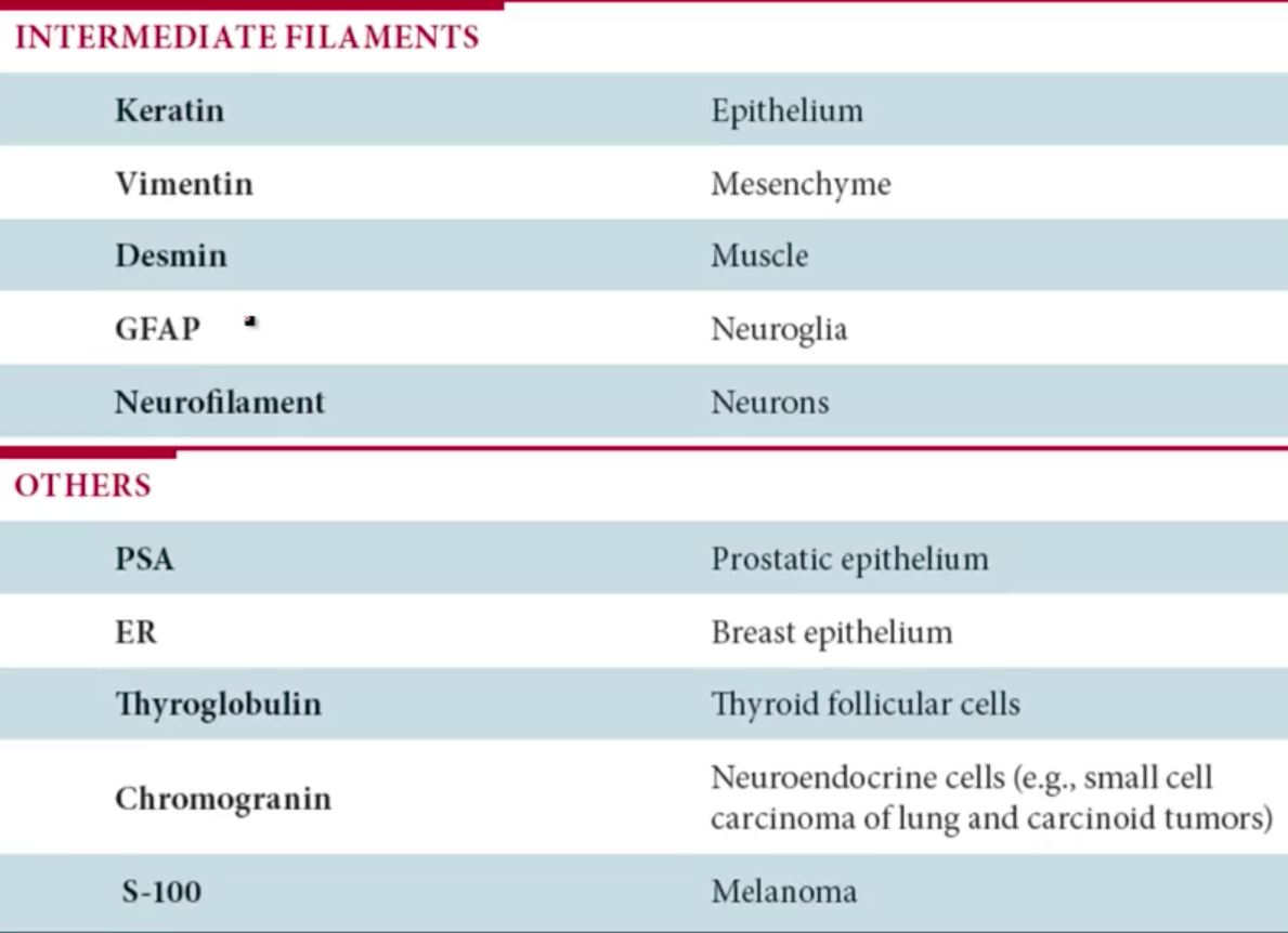
intermediate filaments of cells differ in different types of cells
epithelium: all intermediate filaments in this type of cell is keratin
mesenchyme: CT, e.g. sarcoma
Physiology
MMSE
_..
concentration: counting months backwards
comprehension: follow commands
Inflammation Mediators
_..


Microbiology
Diphtheria
_..
Toxin penetration through the blood-nerve barrier, leading to peripheral neuropathy, occurs with diphtheria.
Fibrinous exudate → systemic circulation → cortical neurons: This is the route that diphtheria toxin takes from the pseudomembranous exudate it forms in the pharynx to the bloodstream and subsequently to cardiac and cerebral cortical tissues.
Pharm
Zileuton
_..
Zileuton is a selective inhibitor of the lipoxygenase pathway that leads to decreased formation of leukotrienes. Zafirlukast and montelukast are leukotriene D4 receptor antagonists. These agents are typically used for chronic asthma prophylaxis.
Levodopa
Long-term treatment of Parkinson disease (PD) with levodopa can be complicated by periodic fluctuations in motor function: patients have good mobility during "on" periods and increased bradykinesia/rigidity during "off" periods ("on-off" phenomenon). Oftentimes, these fluctuations appear to correlate with serum drug levels (eg, reduced mobility 4 hours after the last dose). As PD progresses, the therapeutic window for levodopa narrows, possibly due to natural or levodopa-induced nigrostriatal degeneration. As a result, small changes in serum drug levels can result in motor fluctuations (eg, mild elevations may result in dyskinesia, whereas slight reductions may result in bradykinesia/rigidity). In advanced PD, motor fluctuations can occur independently of medication dosing and may become unpredictable.
NSAIDS
_..
Over-the-counter analgesics such as nonsteroidal anti-inflammatory drugs (NSAIDs) can cause renal failure (analgesic nephropathy) if taken in large amounts over extended periods. Affected patients typically have a modest elevation in serum creatinine, mild proteinuria, and evidence of tubular dysfunction (polyuria, nocturia). Microscopic hematuria and sterile pyuria may also be seen on urinalysis.
NSAIDs concentrate in the renal medulla along the medullary osmotic gradient, with higher levels in the papillae. These drugs uncouple oxidative phosphorylation and are thought to cause glutathione depletion with subsequent lipid peroxidation, resulting in damage to tubular and vascular endothelial cells. Prolonged use results in chronic interstitial nephritis, seen as patchy interstitial inflammation with subsequent fibrosis, tubular atrophy, papillary necrosis and scarring, and caliceal architecture distortion. Calcium may deposit in areas of chronic inflammation and this calcification is visible on renal imaging. NSAIDs also decrease prostaglandin synthesis, causing constriction of medullary vasa recta and ischemic papillary necrosis.
Vitamin E
_..
Vitamin E is a lipid-soluble, antioxidant vitamin that is widely available in the diet. Vitamin E deficiency is rare but may occur in patients with fat malabsorption and abetalipoproteinemia. The most notable manifestations include hemolysis and neurologic dysfunction (due to free radical damage of cell membranes).
Neurologic symptoms of vitamin E deficiency closely mimic Friedreich ataxia (an autosomal recessive degenerative condition) as many of the same areas in the nervous system are affected. Specifically, patients with either condition may have ataxia (due to degeneration of spinocerebellar tracts), loss of position and vibration sense (due to degeneration of the dorsal columns), and loss of deep tendon reflexes (due to peripheral nerve degeneration). Vitamin B12 deficiency may also present similarly due to subacute combined degeneration of the dorsal and lateral spinal columns.
Down and Heart
_..
Congenital heart defects (eg, complete atrioventricular defect, ventricular or atrial septal defect) are seen in 50% of patients.
Capsaicin
_..
Capsaicin is an irritant found in the chili pepper family. It causes excessive activation of TRPV1 (a transmembrane cation channel), causing a buildup of intracellular calcium that results in long-lasting dysfunction of nociceptive nerve fibers (defunctionalization). In addition, capsaicin causes release and subsequent depletion of substance P, a polypeptide neurotransmitter involved in transmission of pain signals. On initial application, topical capsaicin causes burning, stinging, and erythema, but persistent exposure leads to a moderate reduction in pain over time.
TSST
_Has been associated with the use of tampons and nasal packing.,
_..
Staphylococcus aureus strains producing toxic shock syndrome toxin (TSST) are responsible for most cases of TSS. TSST acts as a superantigen that activates large numbers of helper T cells (compared to a regular antigen, which activates few helper T cells). Superantigens interact with major histocompatibility complex molecules on antigen-presenting cells (eg, macrophages) and with the variable region of the T lymphocyte receptor to cause a nonspecific, widespread activation of T lymphocytes. Activation of T cells is responsible for the release of interleukin (IL)-2 from the T cells and IL-1 and tumor necrosis factor from macrophages. These ILs cause capillary leakage, circulatory collapse, hypotension, shock, fever, skin findings, and multiorgan failure.
Amyloid angiography
_..
Amyloid angiopathy is a consequence of β-amyloid deposition in the walls of small- to medium-sized cerebral arteries, resulting in vessel wall weakening and predisposition to rupture. The disease is not associated with systemic amyloidoses; rather, the amyloidogenic proteins are usually the same as those seen in Alzheimer disease. Amyloid angiopathy is the most common cause of spontaneous lobar hemorrhage, particularly in adults age >60. Hemorrhage tends to be recurrent and most often involves the occipital and parietal lobes. Occipital lobe hemorrhage is typically associated with homonymous hemianopsia; parietal hemorrhages can cause contralateral hemisensory loss. Frontal lobe hemorrhage is less common but may result in contralateral hemiparesis.
_..
Brain arteriovenous malformation is the most common cause of intracranial hemorrhage in children and tends to be a single lesion.
Bronchiolitis obliterans
Chronic rejection affects the small bronchioli producing the obstructive lung disease known as bronchiolitis obliterans. Initially, histopathology shows lymphocytic inflammation and destruction of the epithelium of the small airways. Subsequently, fibrinopurulent exudate and granulation tissue are found in the lumen of the bronchioli, which ultimately results in fibrosis, scarring, and the progressive obliteration of small airways.
Phenotypical Mixing
The acquisition of a new viral surface protein is often all that is necessary for a virus to infect a new type of host cell. In this scenario, avian and human influenza virus particles infect host pig cells; certain progeny avian virus particles obtain some of the surface components (eg, sialic acid receptors) of the human influenza virus, allowing the avian virus to infect human cells. This exchange is an example of phenotypic mixing, which generally occurs when a host cell is coinfected with 2 viral strains and progeny virions contain unchanged parental genome from one strain and nucleocapsid (or envelope) proteins from the other strain. However, because there is no change in the underlying viral genomes (no genetic exchange), subsequent progeny will revert to having only avian influenza type surface proteins and will again be noninfectious to human epithelium.
NF1
_..
Cutaneous neurofibromas usually manifest during early adolescence as multiple, raised, fleshy tumors (<2 cm) that often increase in size and number with age. These are benign nerve sheath neoplasms predominantly comprised of Schwann cells, which are embryologically derived from the neural crest.
Reassortment
_..

Influenza viruses (orthomyxoviruses) are respiratory pathogens that can affect several species, including humans, birds, and swine. They possess the surface proteins hemagglutinin (HA) and neuraminidase (NA), which are virulence factors required for infectivity. These proteins are also targets of the immune system, and neutralizing antibodies against them can confer immunity to specific influenza strains. As such, HA and NA are under constant selective pressure both to maintain species-specific virulence and evade immune recognition.
Orthomyxoviruses contain a segmented genome, and HA and NA are coded by separate RNA segments. This allows for genetic reassortment when 2 distinct strains infect the same cell. For instance, avian coinfection with a human influenza A virus (which can also infect birds, the reservoir species for all influenza A subtypes) and an animal influenza A virus can lead to the human-type HA and the animal-type NA being packaged together into the same virion. This has the potential to create a novel strain of virus to which humans are susceptible but haveno immunologic resistance. This phenomenon is known as antigenic shift, and is responsible for the majority of pandemics and epidemics of influenza A.
Transformation
_..

Certain strains of Streptococcus pneumoniae express capsular polysaccharides that inhibit phagocytosis, making it a successful pathogen. Strains lacking the capsule are not pathogenic; however, S pneumoniae is able to obtain new genetic material from the environment that is released following the death and lysis of neighboring bacterial cells. This process, known as transformation, allows the bacterium to take up exogenous DNA fragments, integrate the DNA into its genome, and express the encoded proteins. Through this method, nonvirulent strains ofS pneumoniae that do not form a capsule can acquire the genes that code for the capsule and therefore gain virulence.
Bacteria that have the innate capacity to undergo transformation are said to be naturally competent and include Haemophilus, Streptococcus, Bacillus, and Neisseria species.
Insulin
_..

The insulin receptor (IR) is a tetrameric structure consisting of 2 alpha and 2 beta subunits. The alpha subunits are extracellular and provide the binding site for insulin. The beta subunits are intracellular and contain tyrosine kinase domains that are activated when insulin attaches to the alpha subunits. A series of downstream signaling is then triggered, starting with the autophosphorylation of IR, phosphorylation of IR substrates 1 and 2 (IRS-1/2), and ultimately, translocation of glucose transporter-4 (GLUT-4) to the cell membrane.
Tumor necrosis factor alpha (TNF-α) is a proinflammatory cytokine that induces insulin resistance through the activation of serine kinases, which then phosphorylate serine residues on the beta subunits of IR and IRS-1. This inhibits tyrosine phosphorylation of IRS-1 by IR and subsequently hinders downstream signaling, resulting in resistance to the normal actions of insulin. Phosphorylation of threonine residues has similar effects. Catecholamines, glucocorticoids, and glucagon can also induce insulin resistance by this same mechanism.
Benzos
_..

_..
Selective serotonin reuptake inhibitors (SSRIs) are considered first-line therapy for both generalized anxiety disorder and panic disorder. However, SSRIs must be taken for approximately 4 weeks before any noticeable therapeutic effect occurs, and their initial activating effects can lead to increased agitation and anxiety during this period. Therefore, a temporary course of benzodiazepines is sometimes used during SSRI initiation if there is a significant increase in anxiety-related symptoms.
Benzodiazepines are typically used for the treatment of anxiety, insomnia, acute seizures, and alcohol withdrawal. They are divided into 3 categories, short-, intermediate-, and long-acting, based on their half-life (including active metabolites). This patient's increased anxiety is causing insomnia, and prescribing a benzodiazepine would be beneficial. However, she works in a mission-critical position, and minimization of undesirable daytime side effects (eg, fatigue, impaired judgment) is a priority. This is best achieved by prescribing a short- or intermediate-acting benzodiazepine such as lorazepam before bedtime.
Huntington
Huntington disease is an autosomal-dominant neurodegenerative disease caused by an increase in the number of CAG trinucleotide repeats in the gene that codes for the huntingtin protein. Expansion of the protein's polyglutamine region results in a gain-of-function that leads to pathological interaction with other proteins, including various transcription factors.
_..
Transcriptional repression (silencing) is one of the mechanisms by which mutated huntingtin is thought to cause disease. Regulation of transcription occurs in part due to the presence of histones, small proteins that complex with DNA to help compact the strands. Histones can undergo a variety of modifications (eg, methylation, acetylation, phosphorylation) that affect the accessibility of the genome for transcription. Acetylation of histones weakens the DNA-histone bond and makes DNA segments more accessible for transcription factors and RNA polymerases, enhancing gene transcription. In Huntington disease, abnormal huntingtin causes increased histone deacetylation, silencing the genes necessary for neuronal survival. As a result, one of the treatment options under investigation includes histone deacetylase inhibitors that help upregulate survival genes.
Urachal
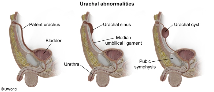
Around 3 weeks gestation, the yolk sac forms a protrusion (allantois) that extends into the urogenital sinus. The upper part of the urogenital sinus gives rise to the bladder. The allantois, which originally connected the urogenital sinus with the yolk sac, becomes the urachus, a duct between the bladder and the yolk sac. Failure of the urachus to obliterate before birth leads to several abnormalities:
Complete failure of obliteration of the urachus results in a patent urachus that connects the umbilicus and bladder. Patients present with straw-colored urine discharge from the umbilicus, which is exacerbated by crying, straining, or prone position. Local skin irritation can cause erythema.
Failure to close the distal part of the urachus (adjacent to the umbilicus) results in a urachal sinus. This presents with periumbilical tenderness and purulent umbilical discharge due to persistent and recurrent infection.
Failure of the central portion of the urachus to obliterate leads to a urachal cyst.
ARF
_..
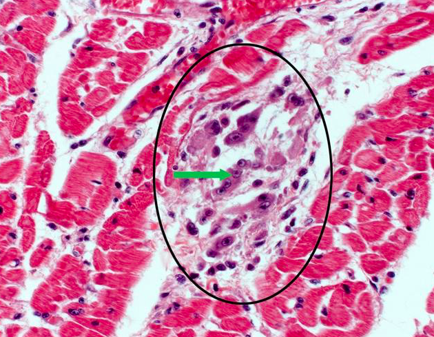
Acute rheumatic fever (ARF) is an immune-mediated complication of an untreated group A streptococcal pharyngeal infection. The most serious manifestation of ARF is pancarditis, which can cause nonspecific fever, fatigue, and anorexia as well as altered vital signs (tachycardia, tachypnea, hypotension). Endocardial involvement resulting in valvular dysfunction (specifically acute mitral valve regurgitation) is the most likely cause of the patient's new holosystolic murmur.
This patient's myocardial biopsy shows interstitial fibrosis with central lymphocytes and macrophages as well as scattered multinucleated giant cells (green arrow). This interstitial myocardial granuloma, or Aschoff body(encircled), is pathognomonic for ARF-related myocarditis. Plump macrophages with abundant cytoplasm and central, slender chromatin ribbons called Anitschkow (or caterpillar) cells are also often present. Over subsequent years, Aschoff bodies are replaced by fibrous scar tissue, leading to chronic mitral valve stenosis and regurgitation.
Inorganic Dust
_..
Inorganic dust is constantly being inhaled and must be cleared by the respiratory tract to prevent disease. The clearance mechanisms used by the lung vary depending on the size of the particles. Larger particles become trapped by mucous secretions in the trachea, bronchi, and proximal bronchioles; these trapped particles are swept upward toward the pharynx by the collective beating of ciliated cells. The finest particles (<2 µm) reach the respiratory bronchioles and alveoli, where they are phagocytized by alveolar macrophages.
Engulfment of inorganic dust causes macrophage activation and release of a number of cytokines that induce pulmonary inflammation. Growth factors, including platelet-derived growth factor and insulin-like growth factor, are also released, which stimulate fibroblasts to proliferate and produce collagen. This production results in progressive interstitial lung fibrosis that characterizes the pneumoconioses.
MAO
_..
The pathophysiologic basis of depression involves the dysregulation of monoamine (eg, serotonin, norepinephrine, dopamine) neurotransmission. Most antidepressive medications act by various mechanisms to increase synaptic concentrations of these neurotransmitters, particularly serotonin. Monoamine oxidase (MAO) is an enzyme located in presynaptic nerve terminals that is responsible for the breakdown of monoamine neurotransmitters. Phenelzine is a monoamine oxidase inhibitor that works by irreversibly binding and inhibiting MAO A and B. This results in increased availability of monoamine neurotransmitters, thereby increasing their release into the synaptic cleft. Because phenelzine irreversibly inhibits MAO, it may take up to 2 weeks following discontinuation of the drug before the enzyme is resynthesized to levels adequate for normal monoamine degradation.
Drug metabolism
The rate and extent of drug metabolism normally varies from person to person. These slight interpersonal variations in the ability to metabolize drugs are typically reflected graphically by a unimodal distribution, usually in the shape of a bell curve, when plasma levels of drug are measured at a fixed time following a fixed dose of drug. This is the method that was used to generate the graph given in the question stem. With most drugs, the majority of people fall within one standard deviation and 95% of people fall within two standard deviations of the population mean of plasma concentration. A single peak in this type of graph indicates that the population being tested possesses a similar genetic drug metabolizing capacity.
A bimodal (discontinuous, polymorphic) curve, as shown in the question stem, results from the presence of two apparently distinct groups within the study population and suggests a pharmacogenetic polymorphism in drug metabolizing capacity. In other words, the two peaks indicate two sets of responders to the drug within the population: one that rapidly converts the drug into its metabolite (considered normals) and another that converts the drug slowly, leading to accumulation of the original drug in the plasma.
Isoniazid is metabolized by acetylation to N-acetyl-isoniazid in the hepatic microsomal system by the enzyme N-acetyl transferase and is subsequently excreted in the urine. The first and second peaks in the above graph represent fast and slow acetylators, respectively. Slow acetylators of isoniazid also metabolize (acetylate) dapsone, hydralazine, and procainamide slowly, causing accumulation of these drugs as well. Slow acetylators of these drugs are at increased risk of toxic effects, while fast acetylators may require much higher therapeutic doses to achieve a therapeutic effect.
(Choice A) Methylation is an important drug biotransformation method to consider when prescribing drugs such as azathioprine and 6-mercaptopurine, drugs used in the treatment of some inflammatory disorders of the bowel and skin.
(Choice B) Glucuronidation is a biotransformation pathway utilized for the metabolism of numerous drugs as well as endogenous substances such as bilirubin. No bimodality has been described with this pathway, but conditions such as Gilbert syndrome involve dysfunction of the glucuronyl transferase system that can lead to toxic accumulation of some drugs.
(Choice C) Hydrolysis occurs with enzymes such as esterases and amidases. Isoniazid is not metabolized by this pathway, and hydrolysis does not exhibit polymorphic metabolism.
(Choice E) Amine oxidation is usually undertaken by monoamine oxidases or by cytochrome oxidative deamination. Neither process metabolizes isoniazid nor exhibits bimodality.
BCR ABL

Approximately four percent of patients with non-small cell lung carcinoma (NSCLC) have an inversion of the short arm of chromosome 2 that creates a fusion gene between EML4 (echinoderm microtubule-associated protein-like 4) and ALK (anaplastic lymphoma kinase). This results in a constitutively active tyrosine kinase that causes malignancy. Interestingly, patients who harbor this gene fusion are usually young non-smokers, often with adenocarcinoma, who lack mutations in either the epidermal growth factor receptor gene or the K-ras gene. The kinase activity of this fusion protein is a target of the protein kinase inhibitor, crizotinib.
The pathophysiology of EML4-ALK NSCLC is most similar to the pathophysiology of chronic myelogenous leukemia (CML). In CML, the classic and most common cause is a translocation between chromosomes 9 and 22. The ABL proto-oncogene is transported from chromosome 9 to chromosome 22 where it is placed adjacent to the BCR gene. The resulting oncogene, BCR-ABL, codes for a fusion protein with constitutive tyrosine kinase activity. This protein stimulates the proliferation of granulocytic precursors and leads to the development of CML. The kinase activity of this fusion protein is a target of the protein kinase inhibitor, imatinib.
Laryngeal
_..
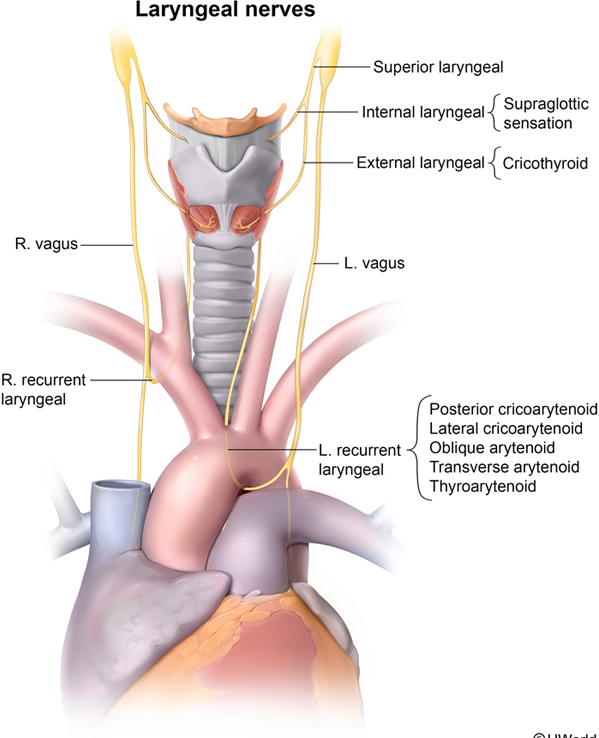
The superior thyroid artery (a branch of the external carotid artery) and the inferior thyroid artery (a branch of the thyrocervical trunk) provide the blood supply to the thyroid and parathyroid glands. The superior thyroid artery and vein and external branch of the superior laryngeal nerve course together in a neurovascular triad that originates superior to the thyroid gland and lateral to the thyroid cartilage. The external branch is at risk of injury during thyroidectomy as it courses just deep to the superior thyroid artery. The cricothyroid muscle is the only muscle innervated by this nerve. It originates on the cricoid cartilage and inserts on the lower border of the thyroid cartilage. The cricothyroid muscle acts to tense the vocal cords, and denervation injury may cause a low, hoarse voice with limited range of pitch.
The internal branch of the superior laryngeal nerve does not innervate any muscles but provides sensory innervation to the laryngeal mucosa above the vocal folds.
_..
IgA antibodies usually bind to pili and other membrane proteins involved in bacterial adherence to mucosa, thus inhibiting mucosal colonization by the microorganism.
Certain bacteria (eg, N gonorrhoeae, N meningitidis, Streptococcus pneumoniae, Haemophilus influenzae) produce IgA proteases that cleave IgA at its hinge region (yielding Fab and compromised Fc fragments), thus decreasing its effectiveness. This facilitates bacterial adherence to mucosa (possibly due to easier bacterial access to mucosal surface or immune disguise by binding to released Fab fragments, among others).
Cushing
_..

This patient's clinical presentation is consistent with Cushing syndrome, the syndrome of glucocorticoid excess. Causes of Cushing syndrome include:
Treatment with pharmacological doses of exogenous glucocorticoids (most common)
ACTH-secreting pituitary adenomas
Ectopic production of ACTH (ie, paraneoplastic)
Primary adrenocortical hyperplasia (eg, adrenocortical tumors)
Of these, only pituitary adenomas and ectopic ACTH production present with elevated ACTH levels.
Transplant rejection

acute rejection: cell mediated
_..
Abduction of the thigh is primarily accomplished by the gluteus medius, gluteus minimus, and tensor fascia lata, which are supplied by the superior gluteal nerve. This nerve exits the pelvis through the greater sciatic foramen above the piriformis.
_..
Extension of the thigh is mostly accomplished by the gluteus maximus muscle, which is supplied by the inferior gluteal nerve. This nerve exits the pelvis through the greater sciatic foramen below the piriformis.
_..
Flexion of the thigh is primarily accomplished by the psoas, iliacus, and sartorius muscles. The psoas is directly innervated by the lumbar plexus, and the iliacus is innervated by the femoral nerve.
A 12-year-old girl is brought to the clinic by her parents after she is found to have hypertension by her school nurse. The patient has no symptoms and reads a book during the office visit. The girl had not received routine well-child care until immigrating to the United States several months ago. She has had several episodes of fever and abdominal pain, which her parents have been able to treat with over-the-counter antibiotics. The patient's blood pressure is elevated on several readings in the office. There is no family history of hypertension. Renal ultrasound reveals dilated calyces with overlying cortical atrophy bilaterally, mostly in the upper and lower poles.
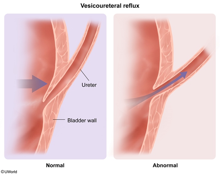
This patient's history of recurrent fever and abdominal pain along with imaging findings consistent with renal scarring indicate recurrent pyelonephritis. Pyelonephritis results from retrograde flow of infected urine from the bladder into the ureter. Normally, the ureters travel through the bladder wall at an oblique angle. When the bladder fills, the intramural ureter becomes compressed. This flap-valve mechanism prevents retrograde flow of urine. However, this mechanism does not work correctly if the ureter enters the bladder wall at a more perpendicular angle, a condition known as vesicoureteral reflux (VUR).
Patients with VUR are at much higher risk for chronic pyelonephritis. Inflammation can occur from pyelonephritis or from VUR itself due to hydrostatic pressure on the papillae. Ongoing injury leads to renal scarring, most commonly at the upper and lower poles of the kidney in which compound papillae are found. Compound papillae are always open, unlike simple papillae in the mid kidney, and are therefore much more susceptible to dilation and subsequent injury. If uncorrected, VUR can lead to loss of nephrons and secondary hypertension.
Posterior urethral valves can present with bilateral hydronephrosis and calyceal dilation due to obstruction of urine flow in the urethra. However, posterior urethral valves result from a malformation of the Wolffian duct, and therefore only occur in males.
_..
Structures arising from neural crest cells are summarized by the mnemonic MOTEL PASS:
melanocytes,
odontoblasts,
tracheal cartilage,
enterochromaffin cells,
laryngeal cartilage,
parafollicular cells of the thyroid,
adrenal medulla and all ganglia,
Schwann cells
spiral membrane.
In emphysema, interalveolar wall destruction decreases the alveolar-capillary surface area, reducing diffusing capacity. Patients with emphysema may have decreased DLCO even when there is little evidence of expiratory airflow obstruction on spirometry.

Excessive bleeding is common in patients with significant renal dysfunction due in part to the accumulation of uremic toxins in the circulation. These toxins impair platelet aggregation and adhesion, resulting in a qualitative platelet disorder characterized by prolonged bleeding time with normal platelet count, prothrombin time (PT), and activated partial thromboplastin time (aPTT). Uremic bleeding can be improved with dialysis as it removes the toxins and partially reverses the bleeding abnormality.
This patient has a nephrotic syndrome, characterized by generalized edema and marked proteinuria (> 3.5 g/day). The presence of nephrotic syndrome and his underlying malignancy suggest membranous glomerulopathy. Membranous glomerulopathy is one of the most common causes of nephrotic syndrome in adults. Up to 85% of membranous glomerulopathy cases are idiopathic. The remainder occur secondary to the following:
Systemic diseases - diabetes mellitus, solid tumors (lung and colon), and immunologic disorders (such as systemic lupus erythematosus)
Certain drugs - gold, penicillamine, and NSAIDs
Infections - hepatitis B, hepatitis C, malaria, and syphilis
This patient's biopsy findings – uniform, diffuse thickening of the glomerular capillary wall on light microscopy without an increase in cellularity (see above image) – are consistent with a diagnosis of membranous glomerulopathy. Electron microscopy reveals that this thickening is caused by irregular, dense deposits laid between the basement membrane and the epithelial cells. These protrusions resemble "spikes" when stained with silver. Immunofluorescence microscopy reveals that these granular deposits contain immunoglobulins (IgG) and C3.
A 30-year-old woman comes to the emergency department with acute-onset shortness of breath. Analysis of the patient’s expiratory gases reveals the following:
Tracheal pO2
150 mm Hg
Alveolar pO2
145 mm Hg
Alveolar pCO2
5 mm Hg
Which of the following best explains the results of this patient's pulmonary gas analysis?

Under normal conditions, the pO2 of inspired air is approximately 160 mm Hg. This value decreases to approximately 150 mm Hg in the trachea due to the partial pressure of water vapor, which proportionately decreases the partial pressures of the atmospheric gases. This patient’s expiratory tracheal pO2 is normal, which indicates that she is breathing room air without any supplemental oxygen.
Normal alveolar pO2 is 104 mm Hg, which lies between the tracheal (150 mm Hg) and venous blood (40 mm Hg) pO2 concentrations. Likewise, normal alveolar pCO2 is 40 mm Hg, again between its respective tracheal (0 mm Hg) and venous blood concentrations (45 mm Hg).
As venous blood flows through the pulmonary capillaries, equilibration occurs with the alveolar gas. Under normal resting conditions, diffusion of both O2 and CO2 across the alveolar membrane is a quick process, with the venous blood only needing to traverse about 1/3 of the total capillary length in order to completely equilibrate. If complete equilibration occurs before the venous blood exits the pulmonary capillary, then equilibration is perfusion-limited. This means that the rate of alveolar capillary perfusion determines the speed with which the alveolar air equilibrates with the venous blood gases. In cases where perfusion is poor, equilibrium will occur slowly or may not occur at all. An example of a severe perfusion defect would be a pulmonary embolus that blocks all blood flow to a given segment of lung that aerates normally. The result is complete failure of atmospheric and venous blood gas equilibration. In this situation, the tracheal air and the alveolar air would have approximately the same composition. The patient described in the question stem has a tracheal pO2 of 150 mm Hg and an alveolar pO2 of 145 mm Hg. Thus, this patient is suffering from very poor alveolar perfusion evidenced by the failure of the alveolar gas to reach its normal equilibrium point.
Aspiration Pneumonia

The cavitary lesion with air-fluid levels described on this patient's chest x-ray is consistent with a lung abscess, which usually occurs as a complication of aspiration pneumonia. Clinically, a lung abscess causes fever, malaise, weight loss, clubbing, and leukocytosis lasting a few weeks. Patients have a cough with copious production of greenish, foul-smelling sputum. Lung abscesses are commonly caused by anaerobic bacteria normally found in the oral cavity or stomach (eg, Fusobacterium, Peptostreptococcus, Bacteroides). Aspiration of the bacteria leads to lung parenchyma necrosis and cavity formation.
Because the right bronchus is straighter than the left, dependent areas of the right lung tend to be affected. If aspiration occurs in the upright position, an abscess in the basal segment of the right lower lobe is likely; if aspiration occurs while supine, the posterior segment of the right upper lobe or the superior segment of the right lower lobe is the most likely location.
Aspiration pneumonia and lung abscesses are associated with:
Altered consciousness (eg, alcoholism, seizures, dementia)
Impaired swallowing (eg, nasogastric tube, dysphagia)
Gingival or upper gastrointestinal tract disease
Lactose Bacteria

The indole positivity (ability to convert tryptophan to indole) of E coli distinguishes it from Enterobacter cloacae, another lactose-fermenting gram-negative rod that is a common cause of UTIs in women
NF1

The MRI image above shows bilateral tumors at the cerebellopontine angle (arrows). Most intracranial schwannomas are found at the cerebellopontine angle and are attached to CN VIII. Schwannomas at this location are called acoustic neuromas. Tinnitus, vertigo, and hearing loss are the typical symptoms of an acoustic neuroma. Bilateral acoustic neuromas occur in neurofibromatosis (NF) type 2. NF-2 differs from NF-1 in that it causes fewer cutaneous manifestations and presents with central nervous system involvement.
Both types of neurofibromatosis are autosomal dominant conditions, although they occur due to mutations on different chromosomes. The difference between these 2 diseases is summarized below.

IVH
_..
A 4-day-old premature infant in the neonatal intensive care unit develops a decreased level of consciousness and hypotonia. She was delivered vaginally at 30 weeks of gestation and her birth weight was 1200 g (2 lb 10 oz). Physical examination reveals a lethargic infant with a weak and high-pitched cry, prominent scalp veins, and tense fontanels. Cranial ultrasound reveals blood in the lateral ventricles. Which of the following structures is the most likely source of the bleeding?

Intraventricular hemorrhage (IVH) is a common complication of prematurity that can lead to long-term neurodevelopmental impairment. It occurs most frequently in infants born before 32 weeks gestation and/or with birth weight < 1500 g, and almost always occurs within the first 5 postnatal days. IVH in the newborn can be clinically silent or present with an altered level of consciousness, hypotonia, and decreased spontaneous movements. Symptoms of catastrophic bleeding include a bulging anterior fontanelle, hypotension, decerebrate posturing, tonic-clonic seizures, irregular respirations, and coma.
IVH in preterm infants usually originates from the germinal matrix, a highly cellular and vascularized layer in the subventricular zone from which neurons and glial cells migrate out during brain development. The matrix contains numerous thin-walled vessels lacking the glial fibers that support other blood vessels throughout the brain, which contributes to the risk of hemorrhage. It is especially vulnerable to hemodynamic instability as premature infants can have impaired autoregulation of cerebral blood flow. Between 24-32 weeks of gestation, the germinal matrix becomes less prominent and its cellularity and vascularity decrease, reducing the risk of IVH.
Other prematurity complications:
NRDS
PDA
bronchopulmonary dysplasia
IVH
necrotizing enterocolitis
retinopathy
Tracheal Deviation
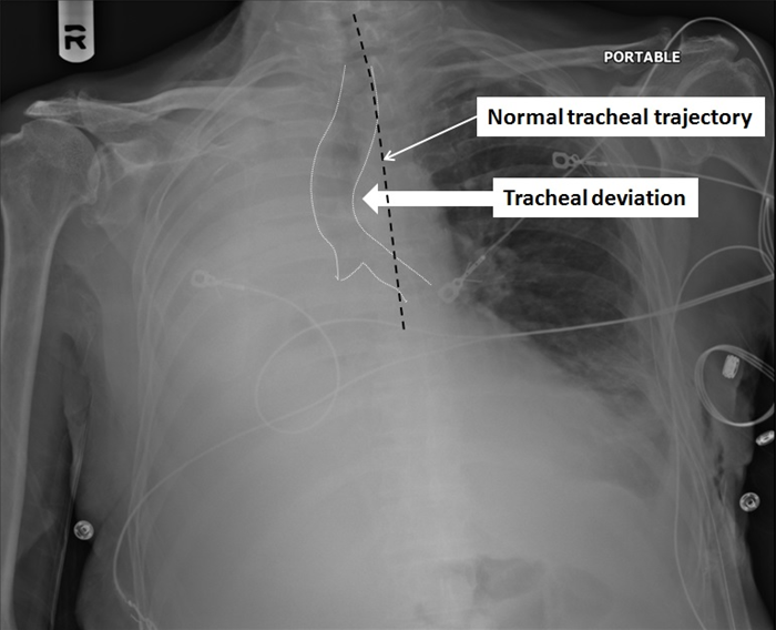
This patient's decreased breath sounds, hemithorax opacification on the right, and deviation of the trachea toward the opacified side are suggestive of a collapsed lung due to bronchial obstruction. Complete collapse of a lung usually occurs following obstruction of a mainstem bronchus (eg, central lung tumors in chronic smokers). As the air trapped in the lung gradually gets absorbed into the blood, there is loss of lung volume due to alveolar collapse (ie, atelectasis), which causes the trachea to deviate toward the affected side. Other mediastinal structures (eg, heart, esophagus, great vessels) may also shift in the same direction. The loss of radiolucent air, combined with shifting of organs into the hemithorax, appears as a completely opacified hemithorax on chest x-ray.
Bookmark
Nigrostriatal degeneration in Parkinson disease results in excessive excitation of the globus pallidus internus by the subthalamic nucleus, which in turn causes excessive inhibition of the thalamus. Reduced activity of the thalamus and its projections to the cortex consequently result in rigidity and bradykinesia. Patients with medically intractable symptoms of Parkinson disease may benefit from high-frequency deep brain stimulation of the globus pallidus internus or subthalamic nucleus. High-frequency stimulation inhibits firing of these nuclei. This causes increased activity in the downstream nuclei, resulting in thalamo-cortical disinhibition with improved mobility.
Keloids and hypertrophic scars consist of connective tissue rich in fibroblasts, myofibroblasts, and bundles of collagen fibers that are arranged in a disorganized fashion (black arrow) in keloids and in parallel in hypertrophic scars.


This patient who recently underwent a genitourinary procedure (cystoscopy) has a urinary tract infection caused by *Enterococcus*. This organism has a morphology of gram-positive cocci in pairs and chains and, when grown on blood agar, reveals no hemolysis (gamma hemolysis). Other characteristics of enterococci include pyrrolidonyl arylamidase (PYR) positivity and ability to grow in bile and in 6.5% sodium chloride. They are unable to convert nitrates to nitrites, explaining this patient's negative result on urinalysis nitrite.
Enterococci are part of the normal intestinal flora of humans and animals. Enterococcus faecalis and E faecium are the most prevalent species cultured from humans and can cause urinary tract infection, bacteremia/endocarditis, wound infection, or intraabdominal or pelvic infection in the nosocomial setting. Enterococci have both intrinsic (beta-lactams, macrolides, aminoglycosides, trimethoprim-sulfamethoxazole) and acquired (vancomycin) resistance to antibiotics, making them important nosocomial pathogens.
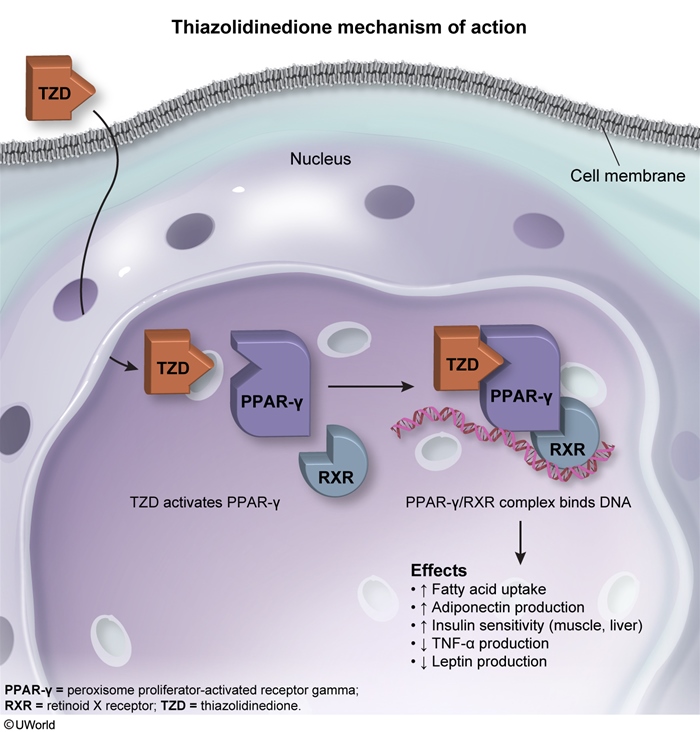
This clinical trial is studying a new thiazolidinedione (TZD), a class of medications that decrease insulin resistance. TZDs (eg, pioglitazone) bind to peroxisome-proliferator activated receptor-γ (PPAR-γ), a transcriptional regulator of genes involved in glucose and lipid metabolism, which subsequently binds additional coactivators. This complex then activates the transcriptional regulatory sequence of the target genes. Important genes upregulated by TZDs include:
Glucose transporter-4 (GLUT4), an insulin-responsive transmembrane glucose transporter expressed in adipocytes and skeletal myocytes that increases glucose uptake by target cells
Adiponectin, a cytokine secreted by fat tissue that increases the number of insulin-responsive adipocytes and stimulates fatty acid oxidation (Choice B)
TZDs are functionally similar to fibrate medications (eg, fenofibrate, gemfibrozil), which bind PPAR-α and have a strong effect in lowering blood triglyceride levels. The PPAR family appears to play a significant role in the pathogenesis of metabolic syndrome (obesity, hypertension, dyslipidemia, and insulin resistance).
Respiratory Epithelium

This patient's verrucous, skin-colored genital lesion is most consistent with condylomata acuminatum, also known as anogenital warts. Condylomata acuminatum is caused by human papillomavirus (HPV), a small double-stranded DNA virus with >100 subtypes. HPV is the most common sexually transmitted disease and can be transmitted by vaginal, anal, and oral intercourse. Most infections are cleared by host immunity, but types 6 and 11 can result in warts.
HPV infects basal epithelial cells through small breaks in the skin or mucosal surfaces. It has a predilection for stratified squamous epithelium, which is found in the anal canal, vagina, and cervix. In the respiratory tract, the true vocal cords are the only area covered with stratified squamous epithelium. The vocal cords undergo near-constant friction and abrasion to produce speech. Stratified squamous epithelium is protective, as deeper cells can replace surface cells that are damaged.
Infants can acquire respiratory papillomatosis via passage through the birth canal of mothers infected with the virus. Warty growths on the true vocal cords can lead to a weak cry, hoarseness, and stridor.

The majority of breast cancer cases occur sporadically in women age >50. Hereditary breast cancer should be suspected in patients diagnosed at a young age, patients with 2 or more primary breast and/or ovarian cancers, and individuals with multiple family members affected by early-onset breast or ovarian cancer. Most cases of hereditary breast and ovarian cancer are associated with mutations in the tumor suppressor genesBRCA1 and BRCA2. Tumor suppressor genes are involved in multiple processes, including:
DNA repair
Cellular differentiation
Checkpoint control of the cell cycle
Transcription factor regulation
BRCA1 and BRCA2 in particular are involved in repair of double-stranded DNA breaks. Mutations in BRCA1 and BRCA2 result in genetic instability, predisposing cells to an increased risk of malignant transformation. Both BRCA mutations are inherited in an autosomal dominantmanner with variable penetrance. Affected women have a 70%-80% lifetime risk for developing breast cancer. Moreover, their lifetime risk of developing ovarian cancer is also increased by up to 40%, although the BRCA2 gene is less likely to lead to ovarian cancer.
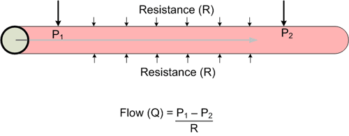
The flow of blood through a vessel can be calculated by dividing the pressure gradient between two points in the vessel by the resistance to flow in that vessel. The resistance in a blood vessel can be calculated using Poiseuille's equation as follows:
Resistance (R) = ηL /r4
η = viscosity of blood L = length of the blood vessel r = radius of the blood vessel (note: raised to the 4th power)
Combining these equations illustrates the profound effect that a small change in blood vessel radius can have on the flow through a vessel. This is due to the fact that the resistance to flow in a blood vessel is inversely proportional to the radius raised to the fourth power.
In the patient described above, flow through the right renal artery has been decreased by a factor of 16 compared to the left renal artery. It can be assumed that the pressure gradient, blood viscosity, and blood vessel lengths are equivalent bilaterally, leaving the blood vessel radius as the only variable. Using the above equation, it can be determined that decreasing the radius by a factor of 2 decreases the flow by a factor of 16.
FLOW = (P1-P2) / ηL r4 = CONSTANT r4, which can be simplified to: FLOW = r4, and: (FLOW / 16) = (r4 / 16) = (r/2)4
Thus, when flow is reduced by a factor of 16, the radius of the lumen is decreased to r/2 (50% of the original radius).
Normal stress vs adjustment disorder with depressed mood:
normal stress, no functional impairment
A 61-year-old man is being evaluated for a 4-month history of easy fatigability and anorexia associated with a 4.5-kg (10-lb) weight loss. Colonoscopy shows a 6-cm mass in the ascending colon. Biopsy samples obtained during the procedure are consistent with adenocarcinoma. A right hemicolectomy is performed. Postoperative evaluation shows that the tumor has invaded several mesenteric lymph nodes and is expressing epidermal growth factor receptor (EGFR). Adjuvant therapy with an EGFR inhibitor is being considered. An activating mutation in which of the following will most likely make the therapy ineffective?

*KRAS* is a proto-oncogene that encodes a GTP-binding protein involved in regulating cell division via transduction of extracellular signals, such as those arising from epidermal growth factor receptor (EGFR). KRAS mutations are found in different types of cancers, including a large proportion of metastatic colon cancers.
Activating mutations in the KRAS gene cause constitutive downstream signaling, even in the absence of EGFR stimulation. This results inincreased cell proliferation and growth that is resistant to anti-EGFR therapy (eg, cetuximab and panitumumab). As such, genetic testing aimed at identifying tumors with wild-type (normal) KRAS or mutated KRAS is recommended prior to initiating therapy.
The dyslipidemia seen with insulin resistance is characterized by high triglyceride and low HDL levels. LDL levels do not increase with insulin resistance.


This patient most likely developed pituitary apoplexy (acute hemorrhage into the pituitary gland), which occurs most often in patients with preexisting pituitary adenomas. Typically, chronic symptoms related to the pituitary tumor (eg, headaches, decreased libido) are present for months before the actual hemorrhage event. The bleeding often presents acutely with severe headache and visual disturbances. Signs of meningeal irritation can also be seen and mimic subarachnoid hemorrhage. However, subarachnoid hemorrhage can be differentiated from a sellar mass with suprasellar extension by the presence of bitemporal hemianopsia, which is present only in the latter. Patients with pituitary apoplexy can develop cardiovascular collapse due to ACTH deficiency and subsequent adrenocortical insufficiency. Pituitary apoplexy is a medical emergency that requires urgent neurosurgical consultation and treatment with glucocorticoids.

The morphologic changes described are consistent with normal aging of the heart. Aging is associated with decreased left ventricular chamber size, particularly in the apex-to-base dimension. This decrease in chamber length causes the ventricular septum to acquire a sigmoid shape, with the basilar portion bulging into the left ventricular outflow tract. Atrophy of the myocardium results in increased interstitial connective tissue, often with concomitant extracellular amyloid deposition. Within the cardiomyocytes, there is a progressive accumulation of cytoplasmic granules containing brownish lipofuscin pigment (the result of indigestible byproducts of subcellular membrane lipid oxidation).
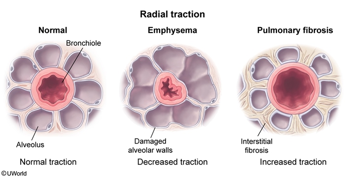
This patient's clinical presentation (progressive dyspnea, fine crackles, clubbing, and diffuse reticular opacities) is consistent with interstitial lung disease. Most interstitial lung diseases cause progressive pulmonary fibrosis with thickening and stiffening of the pulmonary interstitium. This causes increased lung elastic recoil, as well as airway widening due to increased outward pulling (radial traction) by the surrounding fibrotic tissue. The resulting decrease in airflow resistance leads to supernormal expiratory flow rates (higher than normal when corrected for lung volume).
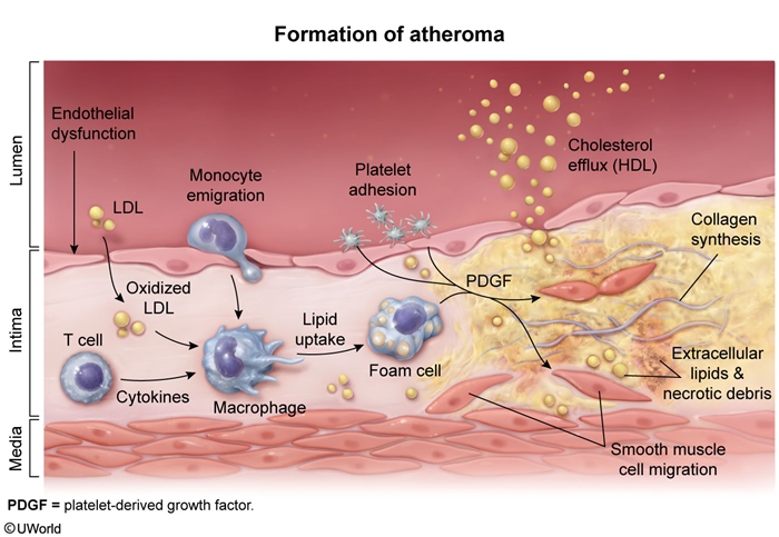
The pathogenesis of atherosclerotic plaques (atheromas) is thought to begin with endothelial cell injury, which results in increased endothelial permeability, enhanced leukocyte adhesion, and altered gene expression. Endothelial dysfunction also promotes platelet adhesion, aggregation, and release of growth factors and cytokines. Platelet-derived growth factor (PDGF) released by locally adherent platelets, dysfunctional endothelial cells, and infiltrating macrophages promotes migration of smooth muscle cells (SMCs) from the media into the intima and increases SMC proliferation. Platelets also release transforming growth factor beta (TGF-β), which is chemotactic for SMCs and induces interstitial collagen production.
Non-Dihydroperidines

The above tracing demonstrates the action potential of cardiac pacemaker cells in cardiac slow-response tissues such as the sinoatrial (SA) and atrioventricular (AV) nodes. The pacemaker action potential includes the following phases:
Phase 4 (spontaneous depolarization) begins after hyperpolarization triggers the opening of HCN channels that allow slow influx of Na+ ("funny current"). T-type (transient) Ca2+ channels then open once the membrane potential becomes more positive, allowing Ca2+ influx to contribute to depolarization. As the pacemaker cell approaches threshold, L-type (long-lasting) Ca2+channels begin to open, which further increases Ca2+ influx and significantly decreases the time until threshold is reached.
Phase 0 (upstroke) is characterized by continued opening of L-type Ca2+ channels. In cardiac slow-response tissues, the action potential upstroke is much slower and transitions gradually from phase 4 due to the relatively slow influx of Ca2+ into the cell.
Phase 3 (repolarization) is characterized by the opening of K+ channels and efflux of K+ from the cell in conjunction with the closure of L-type Ca2+ channels.
This patient's anxiety-provoked episodes of sudden-onset palpitations associated with lightheadedness and shortness of breath are consistent with paroxysmal supraventricular tachycardia. Class IV antiarrhythmics (eg, verapamil, diltiazem) are commonly used to prevent recurrent nodal tachyarrhythmias such as PSVT. They work by blocking L-type calcium channels in cardiac slow-response tissues, which causes slowing of phase 4 depolarization and reduced conduction velocity in the SA and AV nodes.
Ovary

PCP
This man likely experienced substance-induced psychosis from phencyclidine (PCP) intoxication. PCP is a hallucinogen that works primarily as an N-methyl-D-aspartate (NMDA) receptor antagonist; it can work secondarily to inhibit the reuptake of norepinephrine, dopamine, and serotonin. PCP can also have effects on sigma-opioid receptors.
Cell Junctions
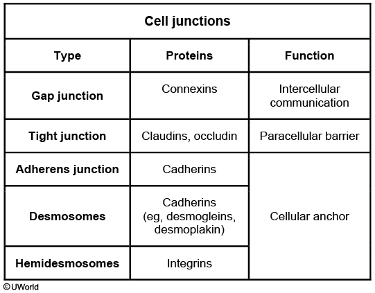
Initiation of labor requires a series of biochemical changes in the uterus and cervix mediated by endocrine changes. Cell junctions are multiprotein complexes that allow contact between adjacent cells or between a cell and the extracellular matrix. Communicating (gap) junctions are especially important during labor and delivery, which require coordination and synchronization of individual myometrial cells. Gap junctions permit diffusion of molecules between neighboring cellular cytoplasms through a connexon (cylinder with a central channel composed of 6 connexin proteins).
Immediately prior to delivery, estrogen stimulates upregulation of gap junctions between individual myometrial smooth muscle cells. An increase in gap junction density at delivery heightens myometrial excitability. Gap junctions consist of aggregated connexinproteins (eg, connexin-43) that allow passage of ions between myometrial cells.
Estrogen also increases expression of uterotonic (eg, oxytocin) receptors, which mediate calcium transport through ligand-activated calcium channels. The combination of an increase in gap junction density and uterotonic receptors results in coordinated, synchronous labor contractions.
Cleft Lip

The lip and palate form during the fifth-sixth week of embryonic development through a series of fusions:
The first pharyngeal arch splits into the upper maxillary prominence and the lower mandibular prominence.
Fusion of the 2 medial nasal prominences forms the midline intermaxillary segment. The intermaxillary segment will become the philtrum of the upper lip, the 4 medial maxillary teeth, and the primary palate.
The left and right maxillary prominences then fuse with the midline intermaxillary segment to form the upper lip and primary palate. If one of the maxillary prominences fails to fuse with the intermaxillary segment, a unilateral cleft lip results. If both maxillary prominences fail to fuse with the intermaxillary segment, bilateral cleft lip results.
Back Pain

This patient has a number of features worrisome for spinal metastases, including advanced age, pain that is worse at night, and persistent and progressive pain that is not relieved with position changes or analgesics. Patients with malignant back pain may also experience systemic symptoms such as fever, weight loss, and night sweats. Known history of malignancy is also a strong predictor of metastatic back pain.
Prostate cancer is the most common malignancy in older men, and it frequently metastasizes to the axial skeleton and proximal femurs. Other malignancies with a propensity for bony metastasis include breast, kidney, thyroid, and lung (PB/KTL: mnemonic "lead kettle").
Headache


Pheochromocytoma

Integrins
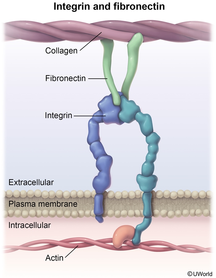
Cellular adhesion is the means by which one cell binds to another cell, surface, or matrix. The integrins are a family of transmembrane protein receptors that interact with the extracellular matrix by binding to specific proteins, including collagen, fibronectin, and laminin. Other adhesion molecule classes include the cadherins, selectins, and Ig superfamily members.
Fibronectins are large glycoproteins produced by fibroblasts and some epithelial cells. Fibronectin binds to integrins, matrix collagen, and glycosaminoglycans, serving as a mediator of cell adhesion and migration. Differential expression of integrin subtypes affects adhesion properties of individual cells, and correlates with malignant behavior in a number of tumors, including melanoma.
Cellular Injury

Imperforate Hymen

An imperforate hymen is an obstructive lesion caused by incomplete degeneration of the central portion of the fibrous tissue band connecting the walls of the vagina. At birth, vaginal secretions stimulated by the mother's estrogen can cause mucocolpos (accumulation of mucus in the vaginal canal), which may manifest as a bulging introitus. If the condition remains undiagnosed, the mucus is reabsorbed and the child will be asymptomatic until menarche. The patient may then present with primary amenorrhea and normal secondary sexual characteristics with cyclic abdominal or pelvic pain due to accumulation of menstrual blood in the vagina and uterus (eg, hematocolpos). The pressure from the resulting collection of blood can also cause back pain and difficulties with defecation. Secondary sexual development is normal as the patient has no chromosomal or hormonal abnormalities. Examination may show a vaginal bulge and/or mass palpated anterior to the rectum.
Renal stones

Renal calculi occur due to an imbalance of the factors that facilitate and prevent stone formation. Increased urinary concentrations of calcium (hypercalciuria), oxalate (hyperoxaluria), and uric acid (hyperuricosuria) promote salt crystallization whereas increased urinary citrate concentration and high fluid intake prevent calculi formation. A high urine citrate concentration has stone-preventing effects as citrate binds to free (ionized) calcium, preventing its precipitation and facilitating its excretion. In fact, potassium citrate is often prescribed to prevent recurrent calcium stones in adults when dietary modifications are unsuccessful.
Conversely, patients with hypocitraturia are at increased risk for calcium oxalate precipitation and stone formation. Hypocitraturia often occurs in the setting of chronic metabolic acidosis (eg, distal renal tubular acidosis, chronic diarrhea) due to enhanced renal citrate reabsorption.
Endometrial Hyperplasia vs endometriosis
Endometrial hyperplasia is characterized by a greater increase in endometrial gland proliferation as compared to stroma. It often presents with irregular, but not painful, menstrual bleeding.
MS Histology
Demyelination with relative preservation of axons
Accumulation of lipid-laden macrophages (containing the products of myelin breakdown)
Astrocytosis (proliferation in response to injury)
Infiltration by lymphocytes and mononuclear cells
Filgrastim
Filgrastim is a granulocyte colony-stimulating factor analog used to stimulate the proliferation and differentiation of granulocytes in patients with neutropenia.
Pacemaker

This patient has a biventricular pacemaker, a device that requires 2 or 3 leads. If 3 leads are used, the first 2 are placed in the right atrium and right ventricle. The third lead is used to pace the left ventricle. Right atrial and ventricular leads are easy to place as they only need to traverse the left subclavian vein and superior vena cava to reach these cardiac chambers. In contrast, the lead that paces the left ventricle is more difficult to position. The preferred transvenous approach involves passing the left ventricular pacing lead from the right atrium into the coronary sinus, which resides in the atrioventricular groove on the posterior aspect of the heart. It is then advanced into one of the lateral venous tributaries in order to optimize left ventricular pacing.
Reflex

EBV Lymphoma
Primary central nervous system lymphoma (PCNSL) is the second most common cause of ring-enhancing lesions with mass effect in HIV. Classically, large, solitary lesions are more likely to suggest PCNSL rather than toxoplasmosis; however, multiple lesions (as seen in this patient) are also common in PCNSL.
PCNSL is a diffuse, large-cell non-Hodgkin lymphoma of B-cell origin that usually occurs as a late complication of HIV infection. Epstein-Barr virus (EBV) is identified in almost all cases. Diagnosis of PCNSL can be made by positive EBV polymerase chain reaction analysis in the cerebrospinal fluid. Definitive diagnosis requires brain biopsy. Although EBV infections in general can be associated with abnormal T-lymphocytes (eg, atypical lymphocytes), T cell infiltration is not commonly seen on brain tissue biopsy of patients with HIV who have PCNSL. T-lymphocytes are the malignant cells in AIDS-associated T-cell lymphoma, not PCNSL (Choice E).
Leukoplakia
Tobacco use can cause leukoplakia, a precancerous lesion that manifests as white patches or plaques on the oral mucosa. In contrast to the white patches caused by thrush, leukoplakia plaques cannot be easily scraped off.
Gynecomastia

Bicuspid Aortic Valve
Bicuspid aortic valves (right and left aortic cusp fusion) occur in approximately 1%-2% of live births, making it one of the most common congenital heart defects. The abnormally shaped valve experiences increased hemodynamic stress, which accelerates the normal aging process and causes premature atherosclerosis and calcification of the aortic valve. These deposits begin accumulating as early as adolescence and lead to aortic stenosis in >50% of affected patients. Symptomatic aortic stenosis develops on average around age 50 (10 years earlier than the average onset of senile calcific aortic stenosis in patients with normal aortic valves).
GLP
Glucagon-like polypeptide-1 (GLP-1) is secreted by intestinal L cells in response to food intake and regulates glucose by decreasing gastric emptying and increasing insulin release. GLP-1 acts through cell surface receptors that are coupled with a G protein-adenylyl cyclase system. Long-acting GLP-1 agonists (eg, exenatide) are second-line agents used in patients with suboptimal glucose control.
Patau
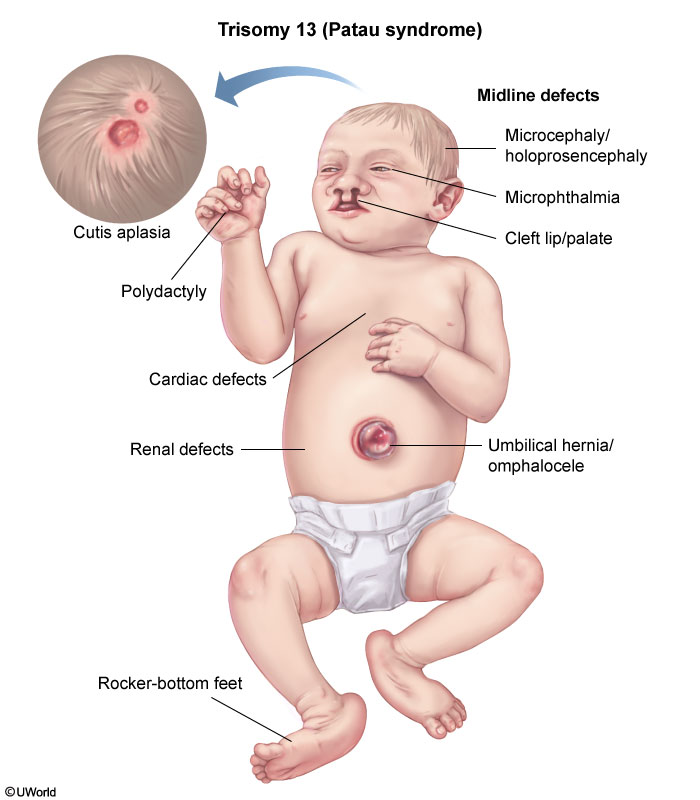
Patau syndrome, or trisomy 13, is a severe genetic disorder with phenotypic features reflecting a defect in the fusion of the prechordal mesoderm, an integral embryological structure affecting growth of the midface, eyes, and forebrain. This results in catastrophic midline defects, including holoprosencephaly, microcephaly, microphthalmia, cleft lip/palate, and omphalocele. Abnormal brain development results in intellectual disability and seizures. Additional abnormalities include polydactyly and cutis aplasia (focal skin defect of the scalp). The majority of patients with Patau syndrome die in utero; only 5% survive beyond 6 months.
Cytogenetic studies usually demonstrate meiotic nondisjunction, which is the failure of chromosomal separation during meiosis, causing inheritance of a chromosome pair from 1 parent rather than a single chromatid. Maternal age ≥35 is an important risk factor for this abnormality of oocyte division. Nondisjunction results in a fetus with 3 complete copies of chromosome 13 (47, XX, +13).
TSST
Staphylococcal Scalded Skin Syndrome (SSSS) is caused by certain strains of Staphylococcus species that produce the exfoliatin exotoxin. Nikolsky's sign (skin slipping off with gentle pressure), epidermal necrolysis, fever and pain associated with the skin rash are the major symptoms of SSSS. SSSS is most common in infants and young children, and it is frequently not fatal unless the skin lesions become secondarily infected.
Exfoliative toxins show exquisite pathologic specificity in blistering only the superficial epidermis ("epidermolytic"). They act as proteases and cleave desmoglein in desmosomes. Bullous impetigo is a more localized form of SSSS with the bulla formation being another effect of exfoliative toxin.
MVP
Primary MVP is most commonly a sporadic disorder and is characterized by myxomatous degeneration (ie, pathologic deterioration of the connective tissue) affecting the mitral valve leaflets and chordae tendineae. Secondary MVP is associated with inherited connective tissue disorders, including Marfan or Ehlers-Danlos syndrome and osteogenesis imperfecta. The myxomatous lesions are characterized by proliferation of spongiosa of the valve leaflets, fragmentation of elastin fibers with increase in mucopolysaccharide, and type III collagen deposits.
Hypothalamic Nuclei

Ear Nerves

The majority of the external ear receives cutaneous innervation from the great auricular nerve, lesser occipital nerve, and auriculotemporal nerve. Most of the external auditory canal, including the external portion of the tympanic membrane, is innervated by the mandibular division of the trigeminal nerve (cranial nerve [CN] V3) via its auriculotemporal branch.
However, the posterior part of the external auditory canal, as well as the concavity and posterior eminentia of the concha, is innervated by the small auricular branch of the vagus nerve (CN X). This patient has experienced vasovagal syncope after stimulation of his posterior external auditory canal by an otoscope speculum. In this form of syncope, parasympathetic outflow via the vagus nerve leads to decreased heart rate and blood pressure.
Medicare
Medicare is a federal socialized medical insurance program that covers select individuals. It provides health insurance for patients age 65 and older who have worked and paid into the system (ie, have paid taxes). Individuals must also hold residence and citizenship in the United States. Medicare also covers younger individuals with disabilities, end-stage renal disease, or amyotrophic lateral sclerosis. Medicare consists of several parts, with Part A covering inpatient hospital visits, Part B covering a select number of outpatient services and medical devices, Part C as an optional capitated plan with additional benefits (vision, dental), and Part D as an optional prescription drug plan. Medicare is different from Medicaid, a state-run medical insurance program that covers the homeless, undocumented immigrants, pregnant women, and low-income families.
In this situation, only the disabled son would be eligible for Medicare.
B2 effect

Papsmear
Clue cells

with small organisms
Actinomyces*-like organisms (black arrow) appear as clusters of basophilic thin filaments that resemble "cotton candy" on a Pap smear; their presence can be an incidental finding in patients with intrauterine devices.

The endometrium consists of simple tubular glands within stroma. These cells resemble histiocytes and have small dark nuclei and no perinuclear clearing. They may be present on a Pap smear during menses. Glandular endocervical cells (black arrow) are columnar cells with a vacuolated or granular cytoplasm and prominent cell borders that form a honeycomb pattern when in clusters. Their presence in a Pap smear indicates adequate sampling.

Maxillary Artery
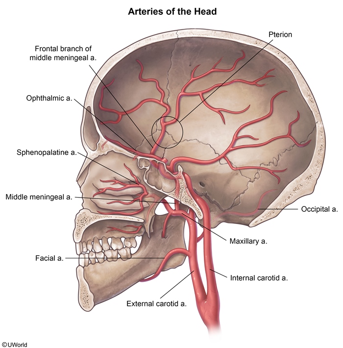
This patient has suffered a fracture at the pterion, a region where the frontal, parietal, temporal, and sphenoid bones meet in the skull. The bone is thin in this region, and fractures there risk lacerating the middle meningeal artery and causing an epidural hematoma. Epidural hematomas require prompt treatment as they are under systemic arterial pressure and can expand rapidly, leading to elevated intracranial pressure (can cause Cushing reflex), brain herniation (eg, uncal herniation with oculomotor nerve palsy), and death.
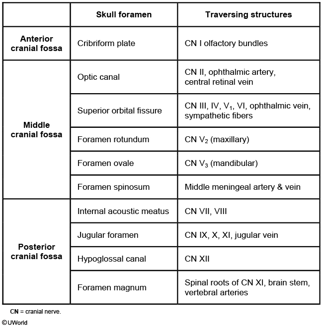
The middle meningeal artery is a branch of the maxillary artery (one of the terminal branches of the external carotid artery), which enters the skull at the foramen spinosum and supplies the dura mater and periosteum.
Kaposi
Kaposi sarcoma is a tumor that results from infection with human herpesvirus 8. It typically manifests as skin nodules but can also affect the gastrointestinal and respiratory tracts. Microscopic evaluation reveals highly vascular tissue containing elongated spindle cells.

Glands
This patient has inflammatory acne. The pathogenesis of acne centers on the sebaceous glands and hair follicles and has 4 major components:
Keratinization of the hair follicle with formation of a keratin plug that blocks release of sebum
Hypertrophy of the sebaceous glands with excess sebum production
Colonization of the glands with Propionibacterium acnes
Bacterial hydrolysis of triglycerides in sebum and release of inflammatory fatty acids
Sebaceous glands are a type of holocrine exocrine gland. Holocrine glands produce their lipid-rich secretory product by lysis of the cell membrane and release of cytoplasmic contents. Other examples of holocrine glands include the Meibomian glands of the eyelid.
The other primary types of exocrine glands include apocrine and merocrine glands. Apocrine glands (eg, mammary glands) produce their secretory product by budding of membrane-bound vesicles. Merocrine glands (eg, salivary glands, eccrine sweat glands) release their watery secretory product via exocytosis, with no loss of the cytoplasmic membrane.
Endocrine glands release hormones into the bloodstream to exert an effect on a remote organ. Paracrine cells produce secretions that reach nearby target cells by diffusion through the extracellular space. Autocrine hormones are also released into the extracellular space but act directly on the secreting cell.
Tuberous Sclerosis
Tuberous sclerosis involves defective tumor suppressor gene-coded proteins hamartin (TSC1) and tuberin (TSC2), and is characterized by cutaneous angiofibromas, brain hamartomas, and cardiac rhabdomyomas.
Neurophysins
Neurophysins are carrier proteins for oxytocin and vasopressin (antidiuretic hormone, ADH), hormones produced within the paraventricular and supraoptic nuclei, respectively, and released from the posterior pituitary. Like the hormones they accompany, neurophysins are produced within the neuronal cell bodies of hypothalamic nuclei. Neurophysins bind oxytocin and vasopressin and act as chaperone molecules as they are shuttled toward the nerve terminals in the posterior pituitary.
Neurophysin II has a binding site specific for vasopressin and is thought to be involved in the transport and packaging of vasopressin through the endoplasmic reticulum (ER) and Golgi apparatus into neurosecretory granules. A point mutation in neurophysin II could result in abnormal protein folding and removal from the ER along with bound vasopressin, thereby decreasing availability of vasopressin for neurosecretory release. Such a mechanism may be responsible for some cases of autosomal dominant hereditary hypothalamic diabetes insipidus.
Drug Reactions

Pseudoallergic drug reactions mimic immunologic reactions but occur via a non-immunologic mechanism (eg, direct immune cell activation, inhibited prostaglandin synthesis). Examples include rhinitis with NSAIDs, anaphylaxis to radiocontrast media, and pruritus with opiates.
Staph Toxins
The preformed heat stable enterotoxin of Staphylococcus aureus can be present most commonly in custard, mayonnaise and processed or salted meats and cause food poisoning leading to abdominal cramping, vomiting and diarrhea. The onset of symptoms is rapid in these cases as the toxin is preformed, and the diarrhea is watery and nonbloody. Symptoms usually resolve within 24 hours.
Thin Basement membrane
Thin basement membrane disease is a disorder of type IV collagen that typically causes asymptomatic microscopic hematuria. The only abnormality is a thin basement membrane.
Phobia treatment
Specific phobia: psychotherapy
social anxiety: propanolol
panic disorder: SSRI
Major Depressive

This patient's month-long history of depressed mood, low energy, loss of interest, insomnia, weight loss, and poor concentration meets the criteria for major depressive disorder. Depressed mood must be present most of the day, almost every day for ≥2 weeks. Appetite, sleep, and motor activity can be increased or decreased. Accurate diagnosis requires ruling out bipolar disorder (no lifetime history of manic or hypomanic episodes) and medical and substance-induced causes. Untreated hypothyroidism is a known medical cause of depression. However, this patient's normal TSH level indicates that her hypothyroidism is well controlled with levothyroxine.
Autonomous
Communicates a choice
Patient able to clearly indicate preferred treatment option
Understands information provided
Patient understands condition & treatment options
Appreciates consequences
Patient acknowledges having condition & likely consequences of treatment options, including no treatment
Rationale given for decision
[P](javascript:deselect(1002))atient able to weigh risks & benefits & offer reasons for decision
Fragile X

MDR1
Human tumor cells have developed the ability to resist chemotherapy in much the same way that many bacteria have developed resistance to antibiotics. One well-studied mechanism by which human tumor cells resist these agents is via the human multidrug resistance (MDR1) gene. The prototype product of this gene is P-glycoprotein, [a transmembrane protein that functions as an ATP-dependent efflux pump](javascript:deselect(1000)). P-glycoprotein is normally expressed in intestinal and renal tubular epithelial cells and functions to eliminate foreign compounds from the body. This protein is also present in the capillary endothelium of the vessels that form the blood-brain barrier. In this location, P-glycoprotein prevents the penetration of foreign compounds into the CNS. In tumor cells, this ATP-powered transmembrane pump protein actively removes chemotherapeutic agents, particularly hydrophobic agents like the anthracyclines.
Drugs such as verapamil, diltiazem, and ketoconazole, among others, have been shown to reduce the action of this multidrug resistance protein in experimental models; the development of agents specifically intended to abolish this mechanism of resistance is currently underway.
Vaccine
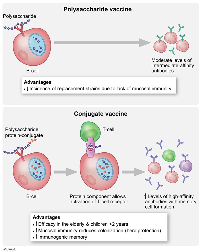
Muscle

The contractile mechanism in skeletal muscle depends on proteins (myosin II, actin, tropomyosin, and troponin) as well as calcium ions. The thick filaments in skeletal muscle are comprised of myosin molecules, with the heads of the myosin molecules forming cross-links with actin during muscular contraction. Two actin chains comprise the thin filaments in skeletal muscle. Tropomyosin molecules sit in the groove between the two actin chains, covering the myosin binding sites on actin when the muscle is at rest. Troponin molecules are small globular proteins situated alongside the tropomyosin molecules. Troponin is composed of three subunits: troponin T, troponin I, and troponin C. Troponin T binds the other troponin components to tropomyosin, troponin I binds the troponin-tropomyosin complex to actin, and troponin C contains the binding sites for Ca2+. During excitation-contraction coupling, Ca2+ is released from the sarcoplasmic reticulum. When Ca2+ binds troponin C, tropomyosin shifts to expose the actin binding sites for myosin, allowing contraction to occur.
Osteoclast

Osteoblasts are cells with a single nucleus that arise from mesenchymal stem cells found in the periosteum and bone marrow. In contrast, osteoclasts originate from the mononuclear phagocytic cell lineage and are ultimately formed when several precursor cells fuse to create a multinucleated mature cell. Osteoclasts in Paget's disease are typically very large and can have up to 100 nuclei (normal osteoclasts have 2-5). The 2 most important factors for osteoclastic differentiation, macrophage colony-stimulating factor (M-CSF) and receptor for activated nuclear factor kappa-B ligand (RANK-L), are produced by osteoblasts and bone marrow stromal cells.
Osteoprotegerin (OPG) is a physiologic decoy receptor that decreases binding of RANK-L to RANK. Inhibition of RANK-L to RANK receptor interaction reduces the differentiation and survival of osteoclasts, resulting in decreased bone resorption and increased bone density (Choice C). OPG loss-of-function mutations cause juvenile Paget's disease. A monoclonal antibody (denosumab) that inhibits the RANK/RANK-L interaction also leads to increased bone density and is commonly used for the treatment of osteoporosis.
Fibroblast growth factors (FGFs) regulate chondrogenesis and osteogenesis. FGFs induce proliferation of osteoblastic precursor cells and anabolic function of mature osteoblasts. Abnormalities in the FGF receptor result in the congenital short-limbed dwarfism known as achondroplasia.
Insulin-like growth factors (IGF-I and IGF-II) are synthesized by various tissues, including the liver and bone. IGF-I increases osteoblastic replication and collagen synthesis; it also decreases collagen degradation by inhibiting the enzyme matrix metalloproteinase-13 (MMP-13). The net effect of IGF-I on the bone is anabolic.
**Transforming growth factor beta increases the replication of osteoblast precursors, leading to increased formation of mature osteoblasts. Transforming growth factor beta also increases collagen synthesis and decreases bone resorption by increasing osteoclastic apoptosis.
Baclofen
Baclofen is a GABA-B agonist that can treat muscle spasticity, which is characterized by increased muscle tone and muscle spasms.
Insurance

Gonadal Arteries

The gonadal arteries arise from the abdominal aorta slightly below the renal arteries. Each gonadal artery courses obliquely downward and laterally within the retroperitoneal space near the psoas major muscle. The right gonadal artery travels in front of the inferior vena cava and behind the ileum, whereas the left gonadal artery courses behind the left colic and sigmoid arteries and iliac colon. After crossing anteriorly over the ureter, the gonadal arteries run parallel to the external iliac vessels and eventually traverse the inguinal canal to supply the testes via the spermatic cord.
Dupuytren
Dupuytren's contracture is a slowly progressive fibroproliferative disease of the palmar fascia. Nodules form on the fascia, eventually resulting in contractures that draw the fingers into flexion. Association with cirrhosis
Hospice Care
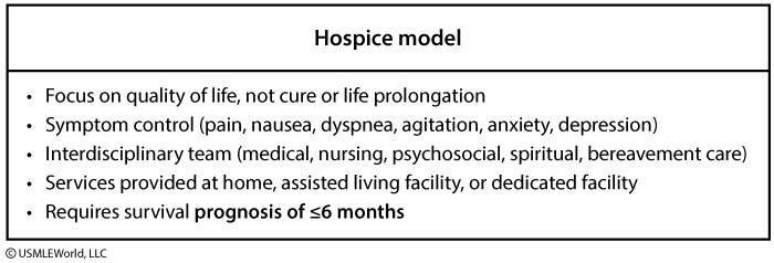
Hospice care is usually provided to terminally ill patients with a prognosis of ≤6 months, when aggressive, curative treatments are no longer beneficial or desired. The largest population receiving hospice care consists of cancer patients, but also includes those with other terminal medical conditions (eg, end-stage cardiomyopathy, end-stage chronic obstructive pulmonary disease, pulmonary fibrosis). The physician must substantiate a prognosis of ≤6 months with documentation of irreversible decline in clinical and functional status.
Ischemic stroke

The coronal section of this patient's brain shows a cystic lesion caused by ischemic stroke involving the left cerebral hemisphere (eg, cortex, subcortical white matter, putamen). Neuropathology coronal sections are oriented with the right side of the brain on the examiner's right side.
Thrombosis of atherosclerotic plaques involving the cerebral arteries can result in vessel occlusion with tissue hypoxia and subsequent necrosis. Ischemic neuronal injury initially causes intense cytoplasmic eosinophilia ("red neurons") followed by neutrophil infiltration a day later. After several days, macrophages/microglia are recruited to phagocytize the necrotic debris and predominate in the ensuing 2-3 weeks. Around this time, the periphery of the necrotic area becomes populated with new blood vessels and proliferating astrocytes that form a network of cytoplasmic extensions (reactive gliosis). Several months to years after brain infarction, the necrotic area appears as a cystic cavity surrounded by a wall composed of dense fibers formed by astrocytic processes (glial scar).
Upper vs Lower

Radial

This patient is exhibiting signs of radial nerve injury (eg, wrist drop). The radial nerve is a terminal branch of the brachial plexus that carries fibers originating in the C5-T1 nerve roots. It innervates most of the forearm extensors at the elbow (eg, triceps) and most of the hand extensors at the wrist. It also innervates the extrinsic extensors of the digits and the brachioradialis and supinator muscles. The radial nerve also provides cutaneous sensory innervation to the dorsal hand, forearm, and upper arm.
Radial nerve deficits in the setting of a midshaft humeral fracture should raise concern for an associated injury to the deep brachial artery. The deep brachial (also termed profunda brachii) artery branches off the brachial artery high in the arm, passes inferior to the teres major muscle, and courses posteriorly along the humerus in close association with the radial nerve.
Waxy Casts
Waxy casts are seen in advanced renal disease (chronic renal failure). They are shiny, translucent tubular structures formed in the dilated tubules of enlarged nephrons that undergo compensatory hypertrophy in response to reduced renal mass.
Osteocytes

Within a single Haversian system, the central canal is encircled by multiple concentric lamellae of bony matrix that each contains lacunae filled with osteocytes and extracellular bone fluid. Delicate canaliculi radiate from each lacuna to create a reticular network with adjacent lacunae, and the cytoplasmic processes of the osteocytes lie within these canaliculi. These cytoplasmic processes send signals to and exchange nutrients and waste products with the osteocytes within neighboring lamellae via gap junctions.
The osteocytes serve to maintain the structure of the mineralized matrix and control the short-term release and deposition of calcium (i.e., calcium homeostasis). The plasma calcium concentration directly dictates the metabolic activity of osteocytes, while parathyroid hormone and calcitonin indirectly influence their metabolic activity. Osteocytes can also sense mechanical stresses and send signals to modulate the activity of surface osteoblasts, thereby helping to regulate bony remodeling.
Cellular Junctions
Tight junctions (zonula occludens) are observed at the apices of glandular cells and consist of two closely adherent cytoplasmic membranes without an intervening space. Tight junctions are the first component of the junctional complex.
(Choice B) Hemidesmosomes are half desmosomes that extend from the basal surfaces of keratinocytes in the stratified squamous epithelium to attach to the basal lamina.
(Choice C) Intermediate junctions (zonula adherens) are a delicate network of cytoplasmic filaments that radiate from the cell membrane to hold adjacent cells together. Intermediate junctions are the second component of the junctional complex.
(Choice E) Desmosomes are small, circular, adherent patches circumferentially placed around cells that comprise the third component of the junctional complex. These patches are particularly common in stratified squamous epithelium and contribute significantly to the structural cohesiveness of tissues subject to mechanical stressors.
Posterior Urethral Valves
Posterior urethral valves in male fetuses can also result in decreased fetal urine output and oligohydramnios.
CF Treatment
Lumacaftor and ivacaftor are CFTR-modulating medications that can potentially help patients with CF by restoring CFTR proteins to the membrane and also by enhancing protein function (eg, chloride transport) at the membrane, respectively. The combination of these 2 medications in patients with homozygous ΔF508 mutations has been shown to improve predicted forced expiratory volume (FEV) and decrease rates of pulmonary exacerbations. G551D mutation
Pulmonary HTN

The chest x-ray in primary pulmonary hypertension reveals enlargement of the pulmonary arteries and right ventricle, which is not evident in this patient.
Nasal bleed

The nasal mucosa is highly vascular and easily irritated by trauma (eg, nose-picking), mucosal dryness, foreign body insertion, and rhinitis (eg, allergy, infection). Epistaxis is very common in children and may be classified as anterior or posterior, depending on the bleeding source. Anterior nosebleeds are by far the most common, and the vast majority occur within the vascular watershed area of the nasal septum (anteroinferior part of the nasal septal mucosa) known as Kiesselbach plexus. Anastomosis of the following vessels occurs in this region:
Septal branch of the anterior ethmoidal artery
Lateral nasal branch of the sphenopalatine artery
Septal branch of the superior labial artery (branch of the facial artery)
Management is directed at stopping the bleeding from Kiesselbach plexus, preferably by direct compression of the nasal alae. Cautery (eg, silver nitrate) of the area surrounding the bleeding site may be necessary for persistent bleeding.
Nasal Drainage
The lateral nasal wall contains the superior, middle, and inferior turbinates (also known as conchae). These 3 bony projections are covered with mucous membrane; they warm, humidify, and filter inspired air and expand and contract in response to environmental changes (eg, temperature, humidity, allergens). The turbinates form corresponding meatuses that serve as drainage pathways. The superior meatus provides drainage for the sphenoidal and posterior ethmoidal sinuses. The middle meatus drains the frontal, maxillary, and anterior ethmoidal sinuses and is the most common site of nasal polyps. The inferior meatus drains the nasolacrimal duct.
Rotator Cuff

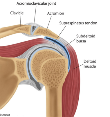
Cyanide Toxicity

This patient's clinical presentation is likely due to cyanide toxicity from nitroprusside infusion. Nitroprusside is a parenteral vasodilator with quick onset/offset mechanics that is commonly used for rapid blood pressure control in patients with hypertensive emergency. It is metabolized in the body to release nitric oxide and cyanide ions.
Cyanide is a potent mitochondrial toxin that binds to Fe3+ in cytochrome c oxidase, inhibiting the electron transport chain and halting aerobic respiration in the cell. Cyanide toxicity presents with altered mental status, seizures, cardiovascular collapse, lactic acidosis, and bright red venous blood (seen on venous blood gas and funduscopy). Nitroprusside-induced cyanide toxicity is most likely to occur in patients receiving higher doses/prolonged infusions or those with renal insufficiency.
Cyanide is normally metabolized in the tissues by rhodanese, an enzyme that transfers a sulfur molecule to cyanide to form thiocyanate, which is less toxic and excreted in the urine. Cyanide overdose depletes the available sulfur donors, allowing cyanide to accumulate in toxic amounts. Sodium thiosulfate works as an antidote by providing additional sulfur groups for rhodanese, enhancing cyanide detoxification. It is used in conjunction with hydroxocobalamin and sodium nitrite in the management of cyanide toxicity.
Ankle

This patient has a right lateral ankle sprain, which is most often due to inversion of a plantar-flexed foot. The ankle is stabilized laterally by the anterior inferior tibiofibular, anterior talofibular, posterior talofibular, and calcaneofibular ligaments (bottom image).
The lateral ankle ligaments are weaker and are injured more often than the medial ligaments. The most common ankle sprains involve only the anterior talofibular ligament and present with pain and ecchymosis at the anterolateral aspect of the ankle. However, stronger forces can injure multiple ligaments, leading to significant joint instability with possible nerve injury and joint dislocation/fracture.
(Choice A) The soleus and gastrocnemius muscles combine to form the Achilles tendon, which inserts on the posterior calcaneus and acts in ankle flexion. The tendon can be injured with sudden forces during strenuous activities (eg, sudden pivoting on a foot or rapid acceleration).
(Choice B) The medial deltoid ligament complex is stronger than the other ligaments and is not commonly injured. Forced eversion of the ankle can damage the deltoid ligament but more commonly causes an avulsion fracture of the medial malleolus.
(Choice C) The subtalar joint is at the posterior junction between the talus and the calcaneus and is reinforced by the talocalcaneal ligaments. The joint is involved with inversion, eversion, dorsiflexion, and plantar flexion of the foot, but is infrequently injured.
(Choice E) Dorsiflexion and/or eversion of the ankle can cause a high ankle sprain affecting the syndesmotic structures (interosseous membrane and anterior, posterior, and transverse tibiofibular ligaments), which connect the tibia and fibula. Injury to these structures is uncommon as they can withstand severe forces. Patients usually have an unstable ankle joint with tenderness at the distal tibiofibular joint, but no significant swelling.
Sleep

Inguinal Ring

Testicles develop in the fetal abdomen during organogenesis. Between 8 weeks and full term, they slowly descend into the scrotum by passing from the abdomen through the deep inguinal ring to enter the inguinal canal. The deep inguinal ring is a physiologic opening in the transversalis fascia bounded by the transversus abdominis muscle laterally and the inferior epigastric vessels medially. The testis then passes anteromedially to exit the canal via the superficial inguinal ring, which is formed by an opening in the external oblique muscle aponeurosis above and medial to the pubic tubercle. Once it has passed through the superficial inguinal ring, the testis enters the scrotum.
Cryptorchidism (failure of one or both testes to descend to the scrotum before birth) affects 3% of males and is more common in preterm infants. A testis that is palpable in the inguinal canal typically descends spontaneously by age 6 months. Testes that have not descended by this point are unlikely to do so and require surgical intervention (ie, orchiopexy). Uncorrected cryptorchidism is associated with a significantly increased risk of testicular cancer and infertility.
In this case, the patient's undescended testicle is lodged within the inguinal canal and must be mobilized through the superficial inguinal ring and stitched into place in the scrotum.
(Choice A) The conjoint tendon is the common tendon of the transversus abdominis and internal oblique muscles. It forms part of the posterior wall of the inguinal canal.
(Choice C) The femoral ring is a physiologic opening between the abdominal cavity and the femoral canal. It transmits lymphatic vessels but not the spermatic cord.
(Choice D) The internal oblique muscle aponeurosis contributes to the formation of the conjoint tendon and rectus sheath. In addition, the cremaster muscle originates from the internal oblique muscle itself.
(Choice E) The rectus muscle sheath is formed by the confluence of the aponeuroses of the external and internal oblique muscles and the transversus abdominis muscle.
(Choice F) An opening in the transversalis fascia forms the deep inguinal ring, which is located superior to the mid-inguinal point (midway between the anterior superior iliac spine and the pubic tubercle). This patient's testis is already in the inguinal canal as it is palpable medial to the deep inguinal ring.
Femoral

The CT image reveals a large fluid collection in the right retroperitoneum lying anterior to the psoas muscle. The fluid is isodense with muscle and displaces the right kidney anteriorly. These findings are consistent with a spontaneous retroperitoneal hematoma, most likely secondary to warfarin use. The risk of bleeding while on warfarin therapy is greatest in patients with risk factors such as increased age, diabetes mellitus, hypertension, and alcoholism.
The femoral nerve descends through the fibers of the psoas major muscle, emerges laterally between the psoas and iliacus muscle, and then runs beneath the inguinal ligament into the thigh. Femoral nerve mononeuropathy can occur due to trauma (eg, pelvic fracture), compression from a hematoma or abscess, stretch injury, or ischemia. Patients with femoral neuropathy develop weakness involving the quadriceps muscle group and may have weakening of the iliopsoas with more proximal nerve injuries. They often complain of difficulty with stairs and frequent falling secondary to "knee buckling." On examination, the patellar reflex is generally diminished. In addition, sensory loss over the anterior and medial thigh and medial leg is typical. Acute, severe pain in the groin, lower abdomen, or back may also occur if the neuropathy is caused by a retroperitoneal hematoma.
Salmonella Typhi
Histology: leukocytes with macrophages
Others: leukocytes with neutrophils
Hearing loss

This patient has Ménière disease, a disorder of the inner ear characterized by increased volume and pressure of endolymph (endolymphatic hydrops) that is thought to be due to defective resorption of endolymph. The resultant distension of the endolymphatic system causes damage to the vestibular and cochlear components of the inner ear. Ménière disease is characterized by the following triad:
Low-frequency tinnitus, or ringing, in the affected ear, often accompanied by a feeling of fullness
Vertigo, the subjective sensation of spinning or motion in the absence of actual motion; commonly associated with lightheadedness, nausea, and vomiting
Sensorineural hearing loss, which is variable in severity but usually worsens over time
(Choice A) Multiple sclerosis (MS) is characterized by patchy demyelination in the central nervous system that varies over time. The presentation of MS is highly variable and may resemble Ménière disease, but most patients will have additional, extra-auditory symptoms (eg, visual symptoms, sensory disruptions).
(Choice C) Labyrinthitis is inflammation of the vestibular nerve that causes acute-onset vertigo, nausea, and vomiting. It usually occurs in a single episode following a viral syndrome.
(Choice D) A mass lesion at the cerebellopontine angle is most commonly an acoustic neuroma (schwannoma of CN VIII). An acoustic neuroma could cause sensorineural hearing loss, vertigo, and tinnitus, but symptoms would be persistent and progressive rather than episodic.
(Choice E) Otosclerosis is an inherited condition seen in middle age. Patients present with conductive hearing loss due to bony overgrowth of the footplate of the stapes. Vertigo does not occur.
HB Traits
However, they may develop hematuria, priapism, and increased incidence of urinary tract infections. Splenic infarction at high altitudes has also been reported.
Polygenic Traits

Hallucinogens
Marijuana THC, cannabis
Increased appetite
Euphoria
Dysphoria/panic
Slow reflexes, impaired time perception
Dry mouth
Conjunctival injection
Vit D
neonates on breastfeed lack
vit D and K (especially if stay inside all the time)
Ureter Blood Supply

The blood supply to the proximal ureter comes from branches of the renal artery. At the distal ureter, arterial blood supply arises from the superior vesical artery. In between, the arterial supply to the ureter is anastomotic and highly variable, with possible afferent branches from the gonadal, common and internal iliac, aorta, and uterine arteries.
Oxyntic
Oxyntic means pink color
Scabies

This patient's presentation – rapidly spreading, pruritic rash with erythematous papules and excoriations on the extremities - suggests scabies. Scabies is due to infestation by the Sarcoptes scabiei mite, which burrows into the skin and spreads through direct person-to-person contact. It usually presents with an intensely pruritic rash in the flexor surfaces of the wrist, lateral surfaces of the fingers, and the finger webs. The rash is often worse at night and is due to a delayed type IV hypersensitivity reaction to the mite, mite feces, and mite eggs. Scabies can also involve other parts of the body (eg, elbows, axillary folds, nipples, and areola in women; scrotum and penis in men), but less commonly affects the back and head (except in children).
Skin examination usually shows excoriations with small, crusted, red papules scattered around the region. Patients can also develop small vesicles, pustules, or wheals. Linear burrows are the most specific finding in scabies, although they are often obscured by excoriations. Diagnosis is confirmed by skin scrapings from excoriated lesions that show mites, ova, and feces under light microscopy, as shown in the image above.
Koilocytosis
infectious koilocytosis: HPV
Shingles
Shingles is characterized by unilateral burning pain and a papular or vesicular rash in a dermatomal distribution. The lesions coalesce, rupture, crust over, and heal within a few weeks although the discomfort may linger for several weeks.
Light microscopy of a sample from a vesicle base reveals intranuclear inclusions in keratinocytes and multinucleated giant cells(positive Tzanck smear). Skin biopsy would show acantholysis (loss of intercellular connections) of keratinocytes and intraepidermal vesicles.
Neonatal O2
Complications of supplemental O2 therapy include (mnemonic: RIB):
Retinopathy of prematurity: retinal vessel proliferation
Intraventricular hemorrhage
Bronchopulmonary dysplasia: oversimplified alveoli due to impaired development
Cancer Associations

Last updated

