10 Cardiac Ischemia
Ischemic Syndrome
_..
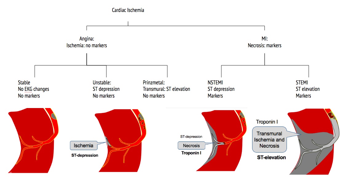
Angina vs MI:
Angina: Symptoms relieved by NG; MI not relieved by NG
stable: unruptured plaque, blocking blood flow. RBC squeeze through. Symptoms with exertion. May have ST depression during symptoms
unstable angina: ruptured plaque, clot, subendothelial (ST depression) ischemia (no markers)
prinzmetal: transmural (ST elevation) ischemia (no markers)
NSTEMI: subendothelial necrosis, clot not completely obstruct artery (not transmural yet)
STEMI: transmural necrosis, clot completely obstructs artery, transmural
_..
coronary ischemia most cause of sudden death, except with HCM in young people
Symptoms
_..
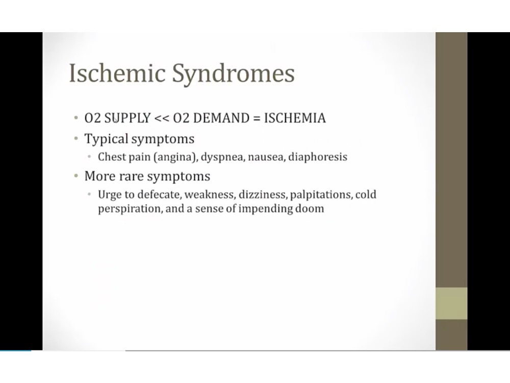
angina: squeezing type of chest pain
lavigne sign: patient place hand over chest
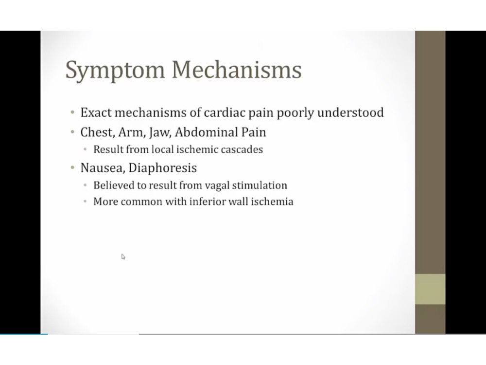
vagus run along inferior wall of heart, can be stimulated with inferior MI
Risks
_..
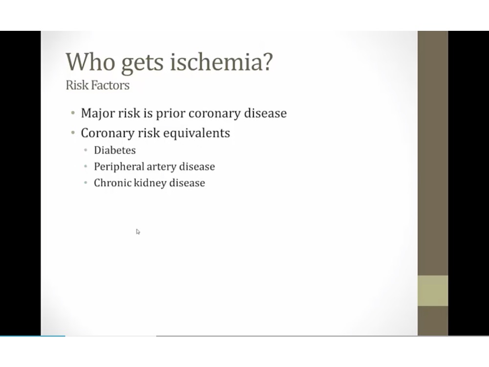
angina, MI in past highest risks
other very high risks
Cardiac Markers
_..
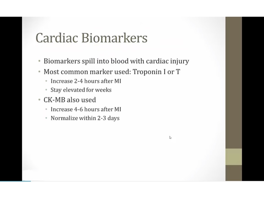
1 hour after chest pain: no cardiac marker, nl biomarkers
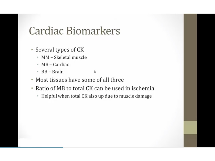
pt with rhabdo will have very high CK, but mostly MM
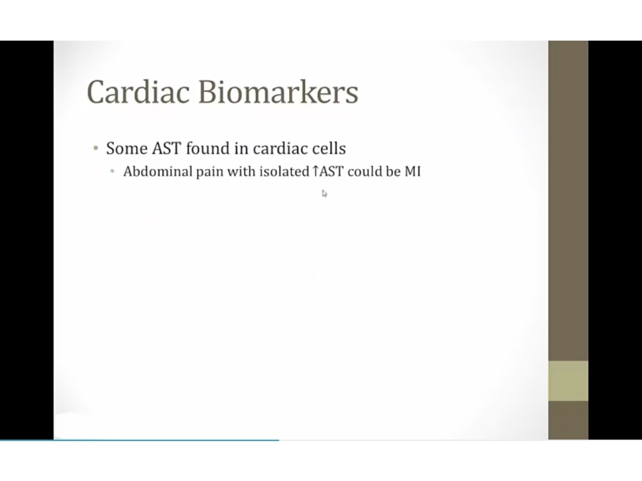
liver enzyme
EKG
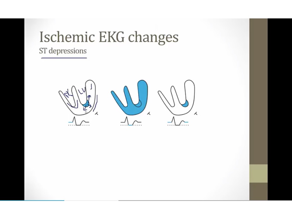
ST depression
_..
blue: small area of subendocardial ischemia
endocardium can get blood from ventricle. Subendo receive blood from epicardial vessels, most vulnerable to ischemia
in mild ischemia: subendo ischemic first
ischemic tissues create electric current going away from it
T: EKG lead looking at LV
At baseline: T sees current heading towards it (elevated ST baseline)
Heart depolarize: everything at normal baseline
repolarize: elevated baseline again
Appears to have ST depression, in reality, baseline elevation
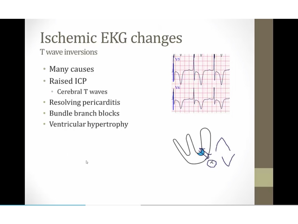
T inversion
_..
normally: subendo repolarizes first, current goes to T, create upward T wave from current heading towards it
subendo ischemia: subendocardium repolarizes last, reversing wave of repolarization, going away from T, inverted T
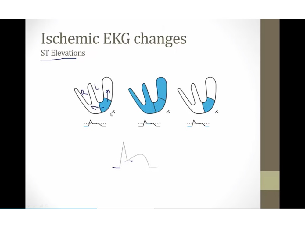
ST Elevation
_..
transmural ischemia, current away from it
baseline: T sees current heading away, depressed baseline
depolarizes: everything at baseline
repolarizes: depressed baseline again
ST elevation = baseline ST depression
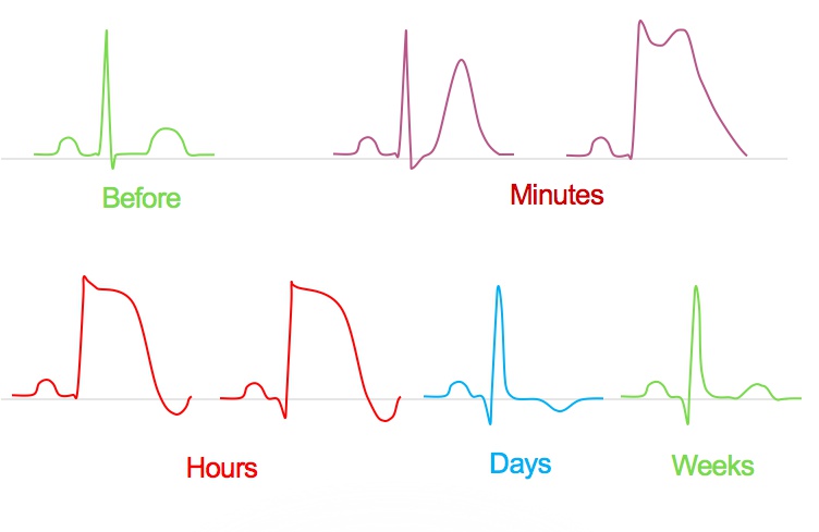
STEMI
_..
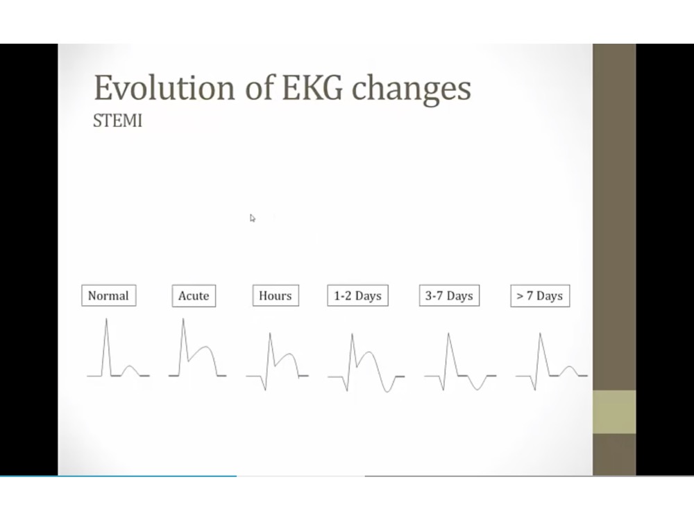
transmural MI progression
Q wave after a few hours, but also represent old infarction
T wave invert after few days
normal ST
T wave normal, Q wave remain
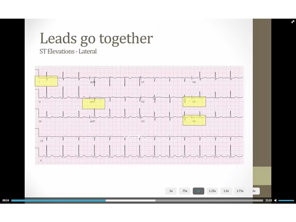
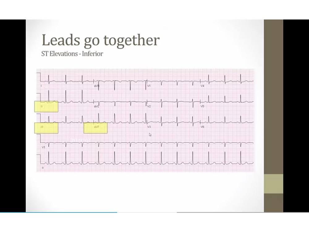
_..
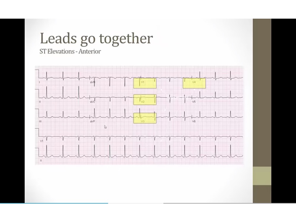
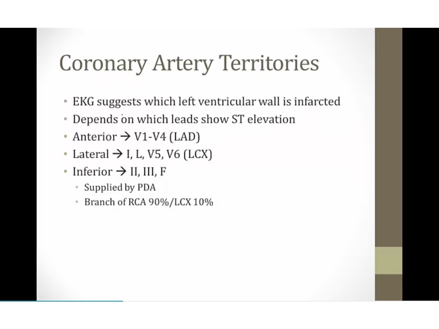
tell which leads ischemic with transmural infarct
anterior wall
Progression and Complications
_..
Overview:
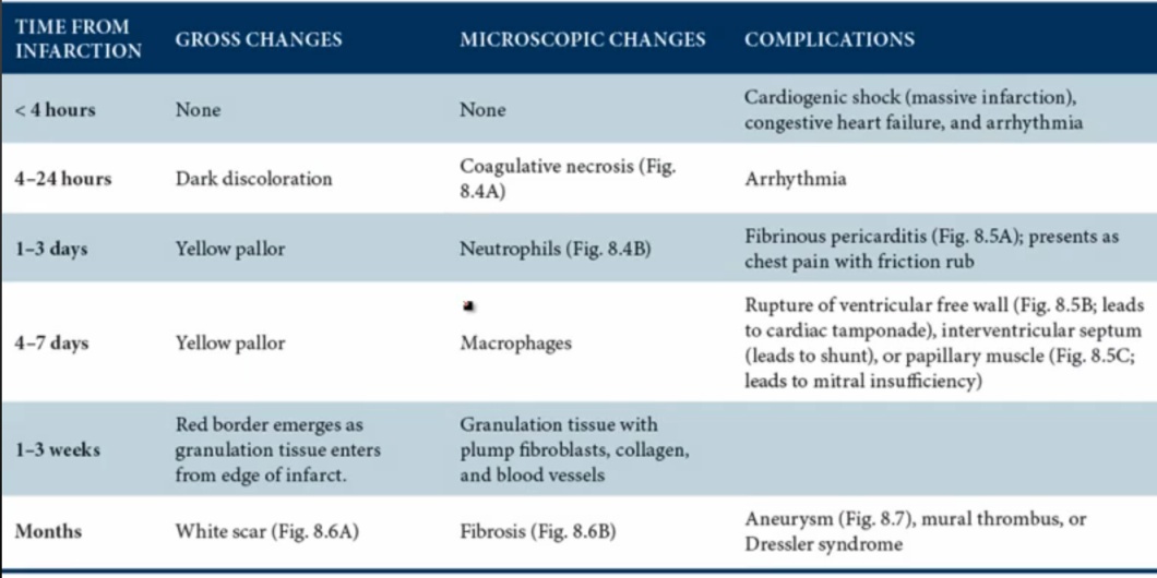
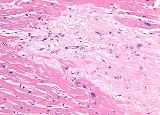
1st thing: coagulative necrosis: removed nucleus from cells
2nd: acute inflammation with neutrophils/macrophages
3rd: healing, granulation, then conversion to scars
< 4 hours:
congestive heart failure: blood back up and can’t pump
arrythmia from damaged conduction
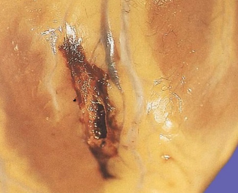
1-7 days:
WBC gives yellow pallor gross color
complication depends on whether neutrophil or macrophages
neutrophil: transmural inflammation, exudate leak to pericardium, pericarditis. Only with transmural inflammation
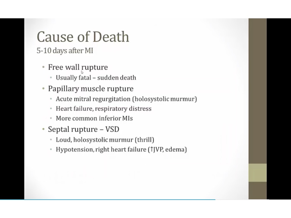
macrophage: eat up all dead debris, wall = weakest, rupture
1-3 weeks:
granulation: blood vessels, red border from outside, from normal tissues
months:
scar: not as strong as myocardium, not good movement, stasis, aneurysm/thrombus
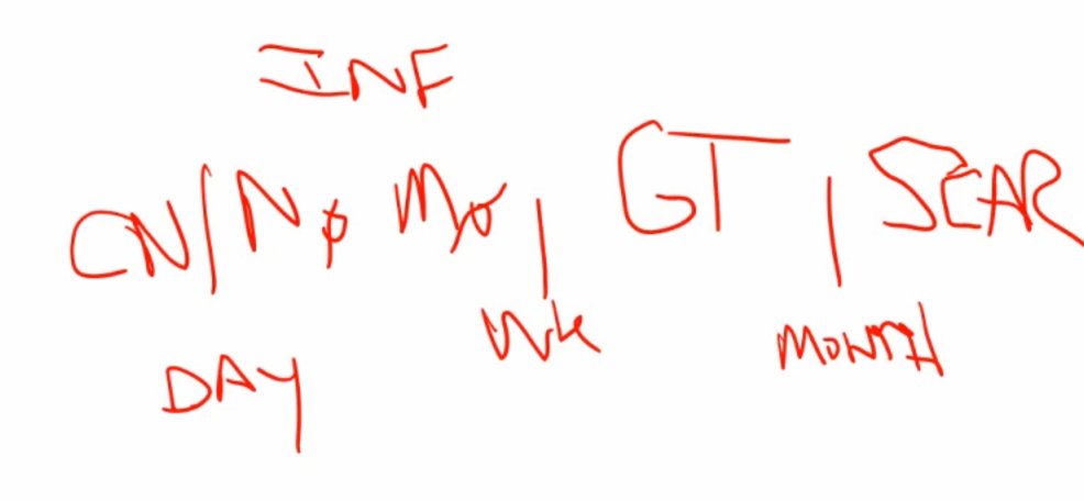
1 day, 1 week, 1 month
1st day: coagulative necrosis
after 1st day: inflammation up to 1 week, neutrophil then macrophage
after 1 week: granulation
1 month: scar
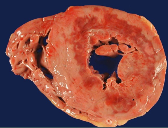
subendo, mottled color
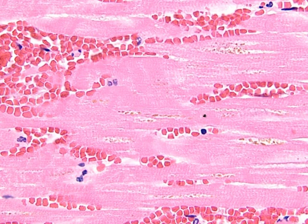
coagulation necrosis
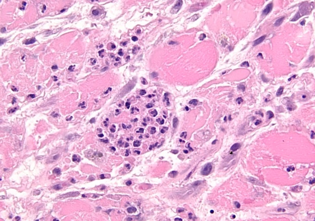
inflammation
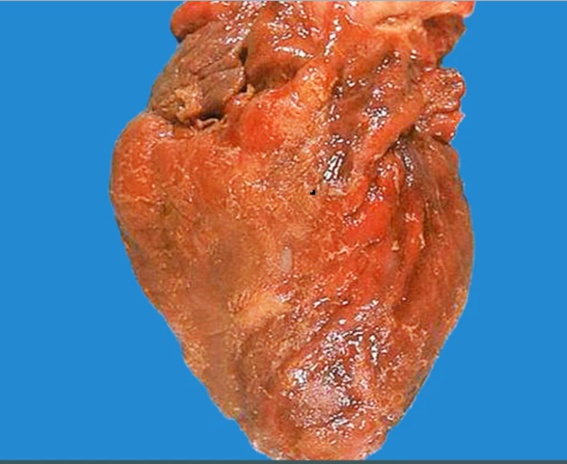
pericarditis
rupture
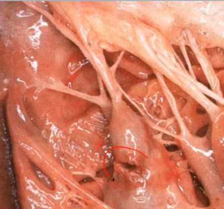
papillary muscle
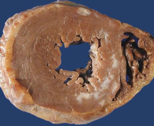
scar
collagen, CT, type 1
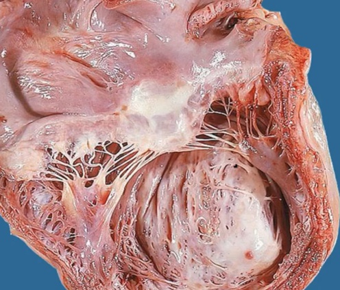
aneurysm
_..
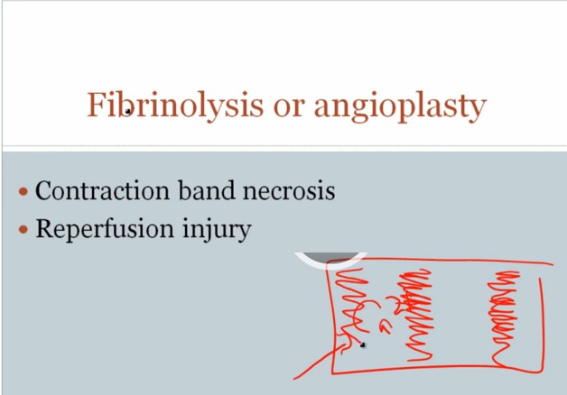
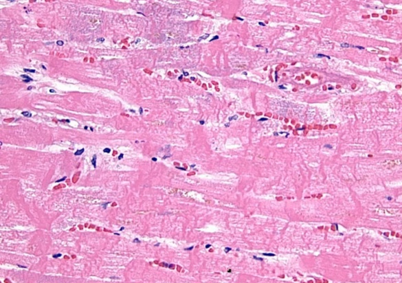
return of blood flow: contraction band
return blood flow, Ca inflow into dead cells, contraction of muscle fibers, dense contraction bands
reperfusion: free radicals from O2 coming back. Cardiac enzyme continue to rise after open up clot
Complications
_..
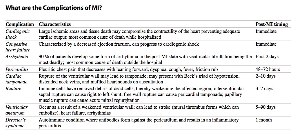
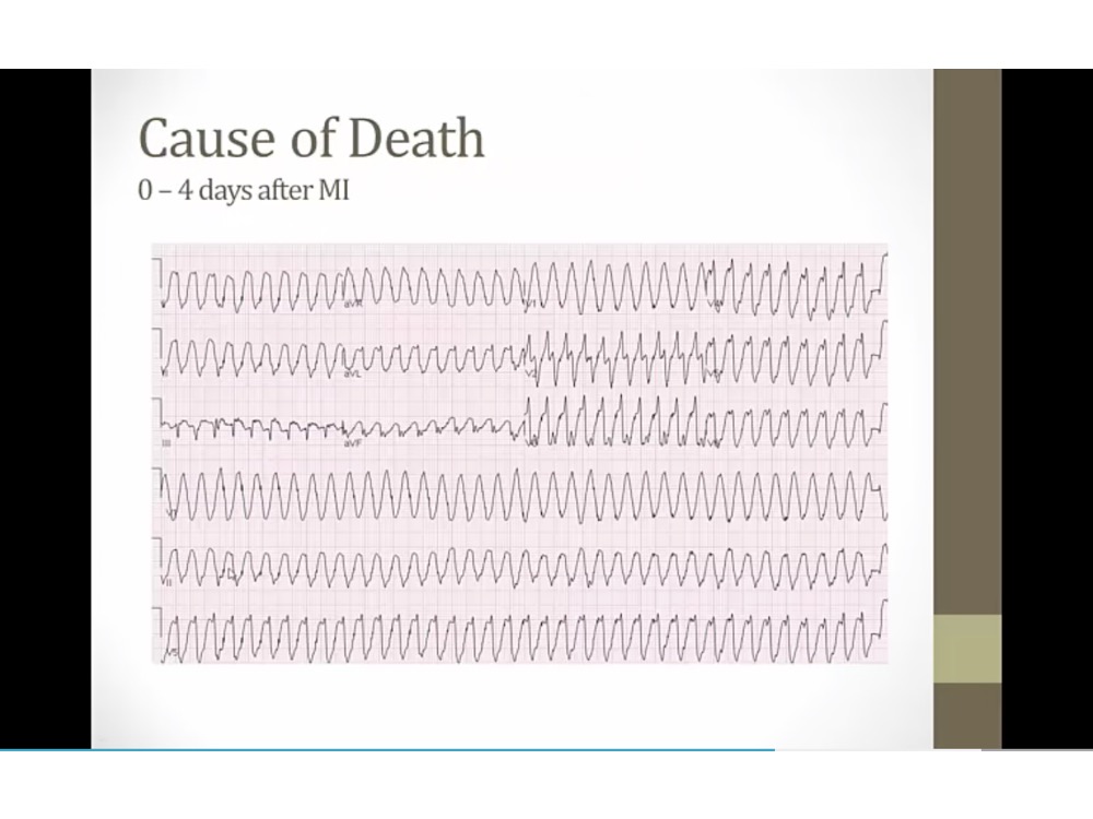
VTACH, can deteriorate into cardiac arrest
can cause tamponade if accumulation of fluid
inferior wall: papillary muscle with single supply from RCA
thrill: feel with hand
hypotension: blood leaks from left to right
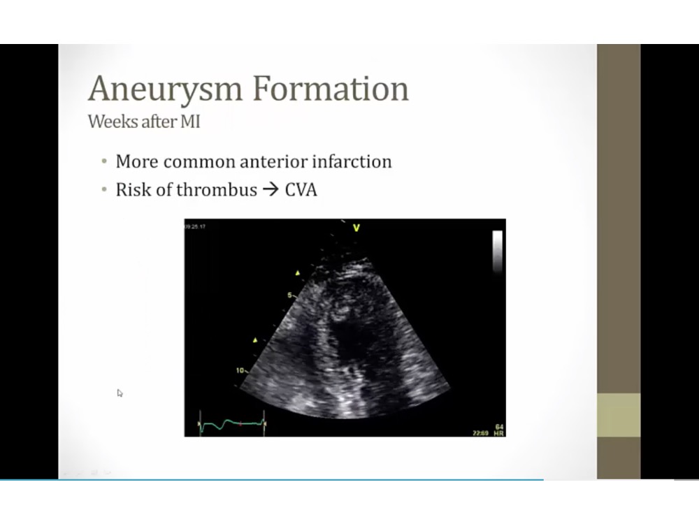
US: apex with akinetic tissue, aneurysm with stasis of blood on left side
can have stroke if a piece breaks off
anterior infection: most common
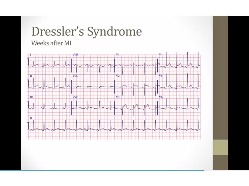
EKG with pericarditis
diffuse ST elevation
PR depression, down going
autoimmune
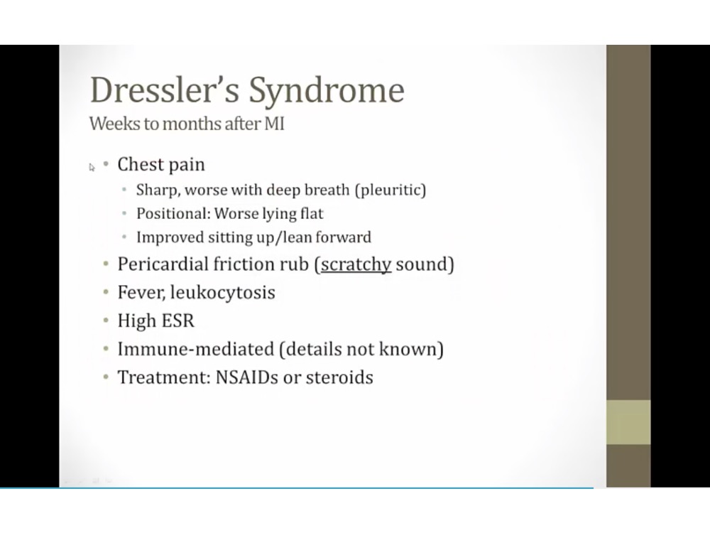
like sand paper
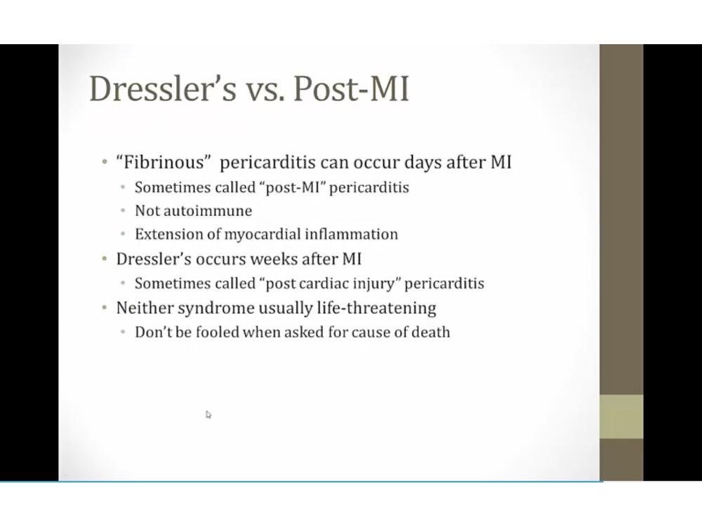
extension of inflammation into pericardium
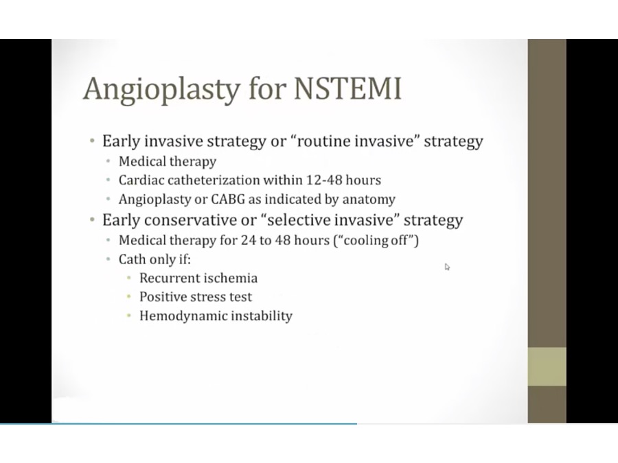
Treatment
Revascularization
_..
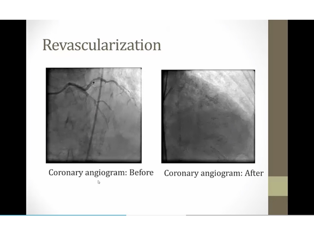
dye with fluoroscope
put in stent
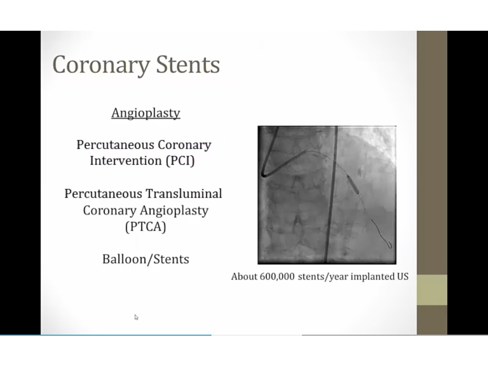
balloon first to push the plaque, then leave stent (chickenwire) in
PCI: going across skin to access artery
PTCA: lumen of artery to get to coronary artery
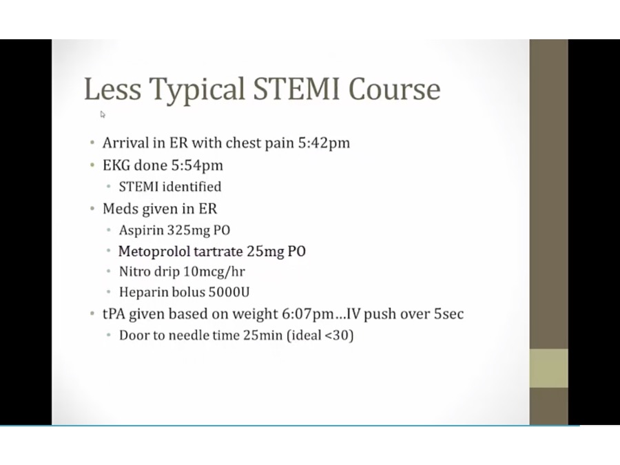
PCI: within 90 min of symptoms onset
more than 90 min: tpa
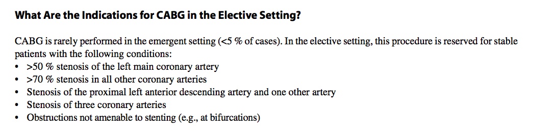
PCI unsuccessful: emergency CABG
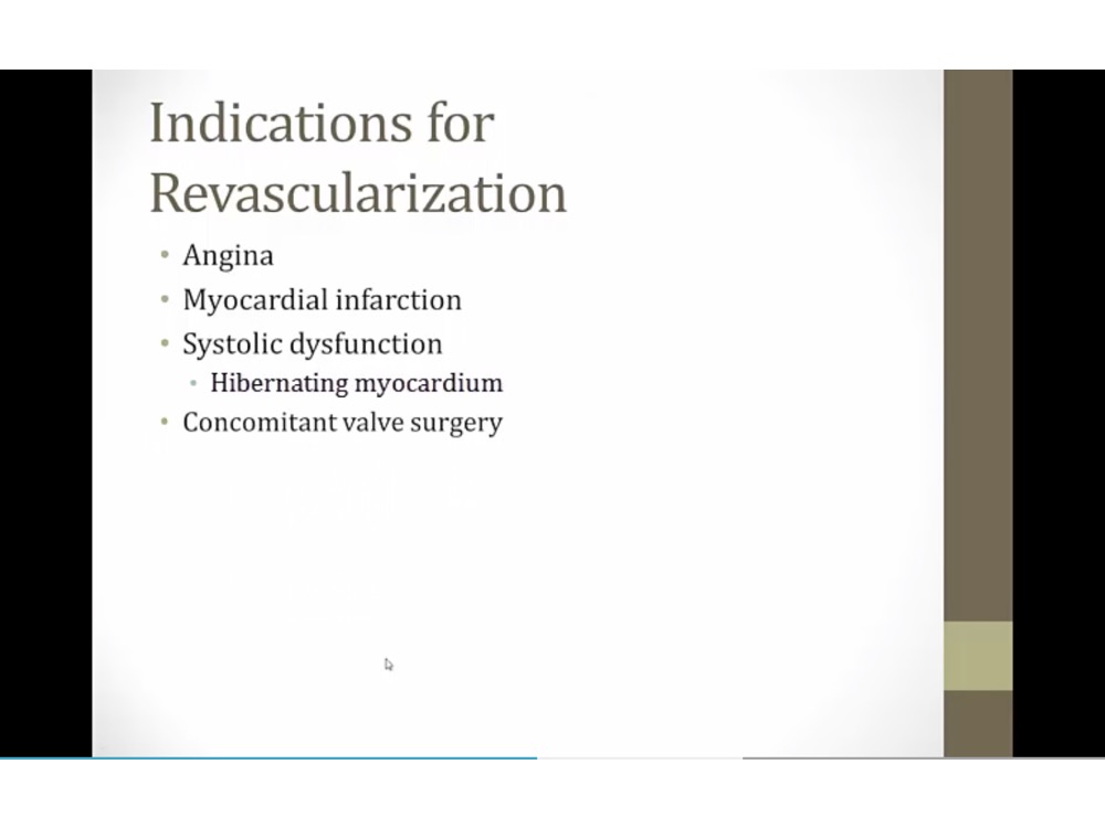
systolic dysfunction: cardiomyopathy with reduced LV EF
hibernating myocardium: myocardium so little flow that going into hibernation
in valve surgery: treat blocked artery at same time
Bypass
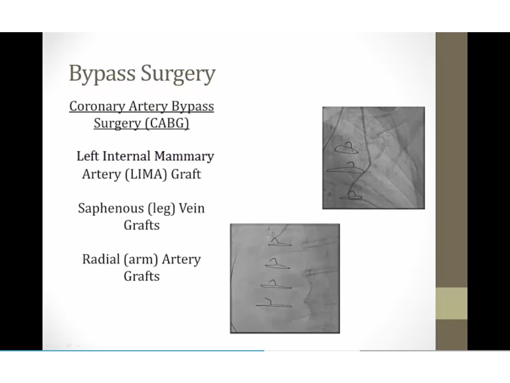
bypass backup option
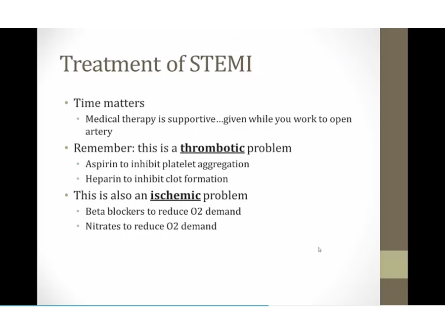
Medication
Prevention
_..
_..
trigger inflammation when put in
drug: sirolimus/tacrolimus to prevent stenosis
thrombosis: complete closure of stent by blood clot inside
endothelialization: scar tissue grew over stent
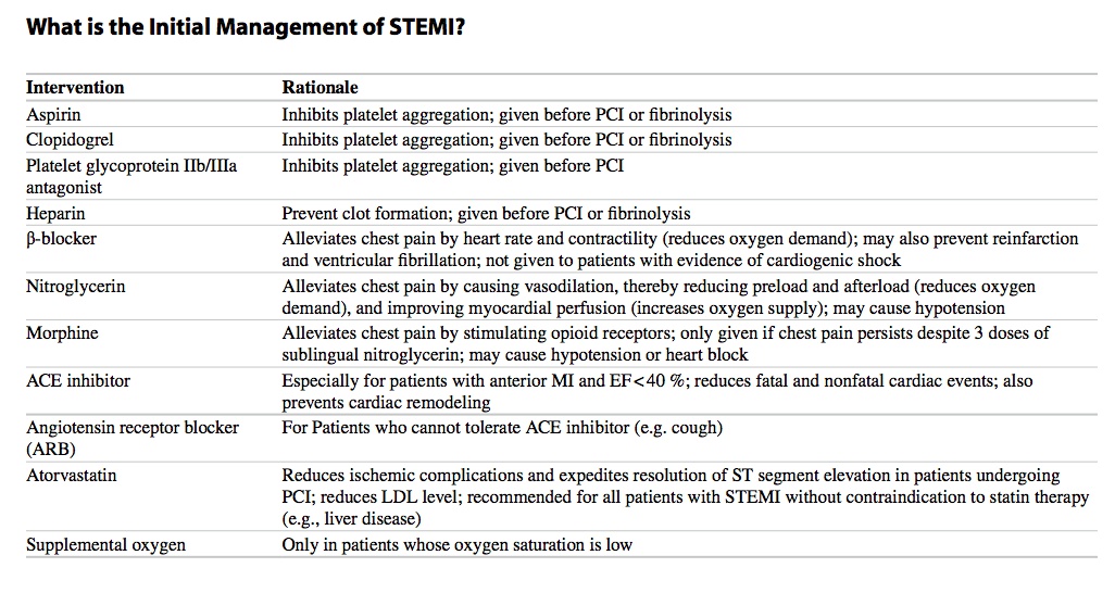
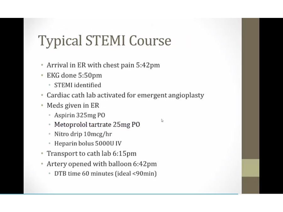
STEMI Treatment
_..
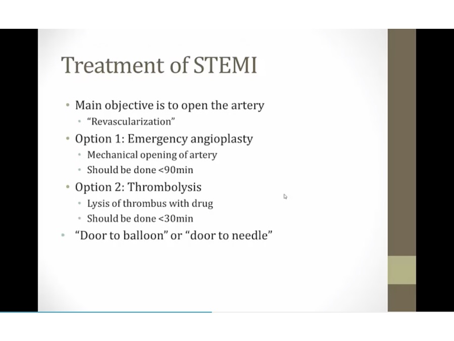
1: catheter
time it takes: door to balloon or needle
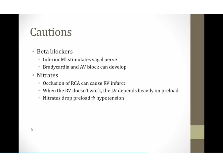
inferior MI: bradycardia and AV block already from parasympathetic stimulation. Adding beta blocker make it worse
usually LV infarct, RV sometimes can be infarcted
hypotension and then cardiac arrest
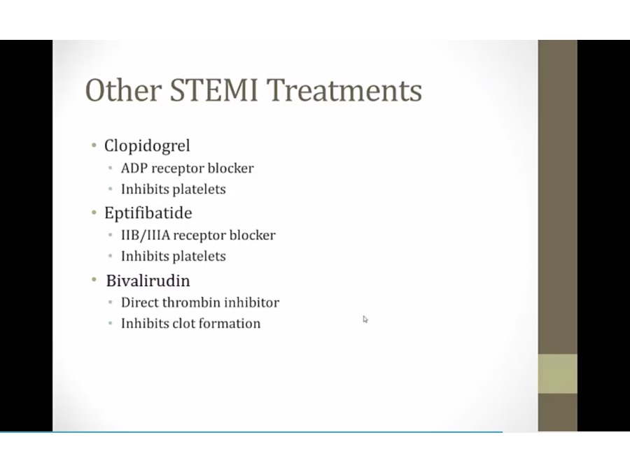

No intRUDIN: bivaliRUDIN is a direct thrombin inhibitor
Big GATOR: arGATROban and dabiGATRAN are direct thrombin inhibitors
ABC sportscaster grabbing fries: abciximab blocks the GP IIb/IIIa receptor preventing platelet aggregation
Antibody-shaped microphones: abciximab is a monoclonal IgG antibody
Tied game: eptifibatide and tirofiban block the GP IIb/IIIa receptor to prevent platelet aggregation
Broken plates: GP IIb/IIIa inhibitors can cause thrombocytopenia
Ketchup time: antiplatelet therapy increases bleeding time (measure of platelet function)
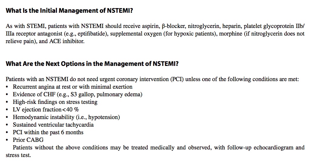
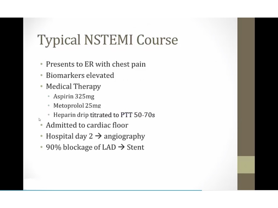
NSTEMI
_..
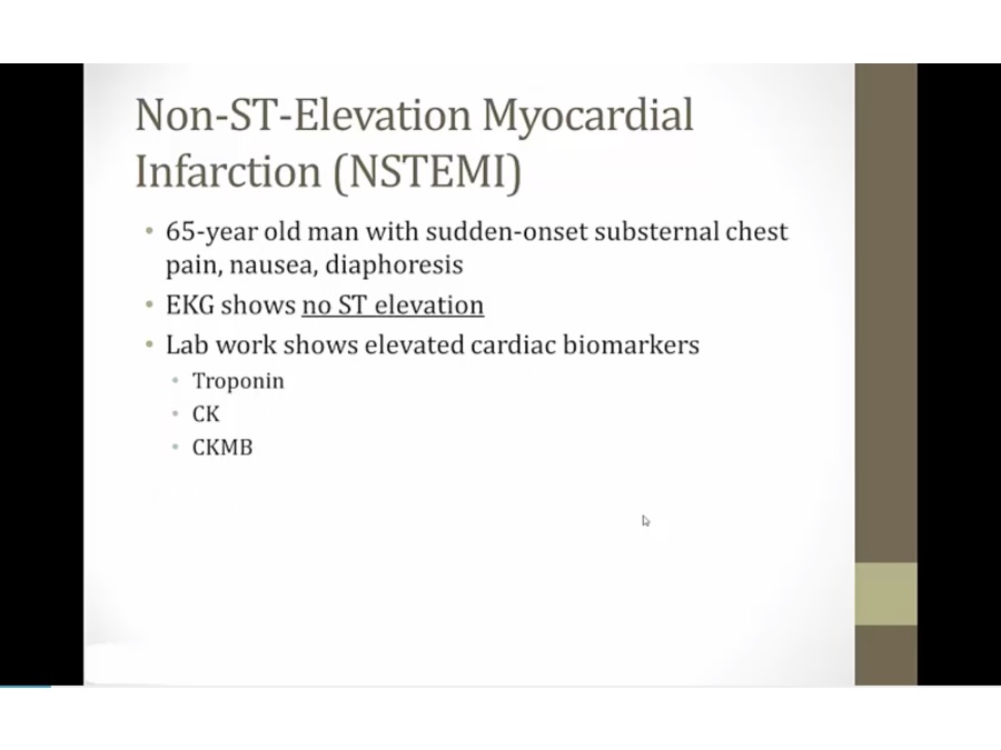
damage to heart tissues but absence of EKG elevation
_..
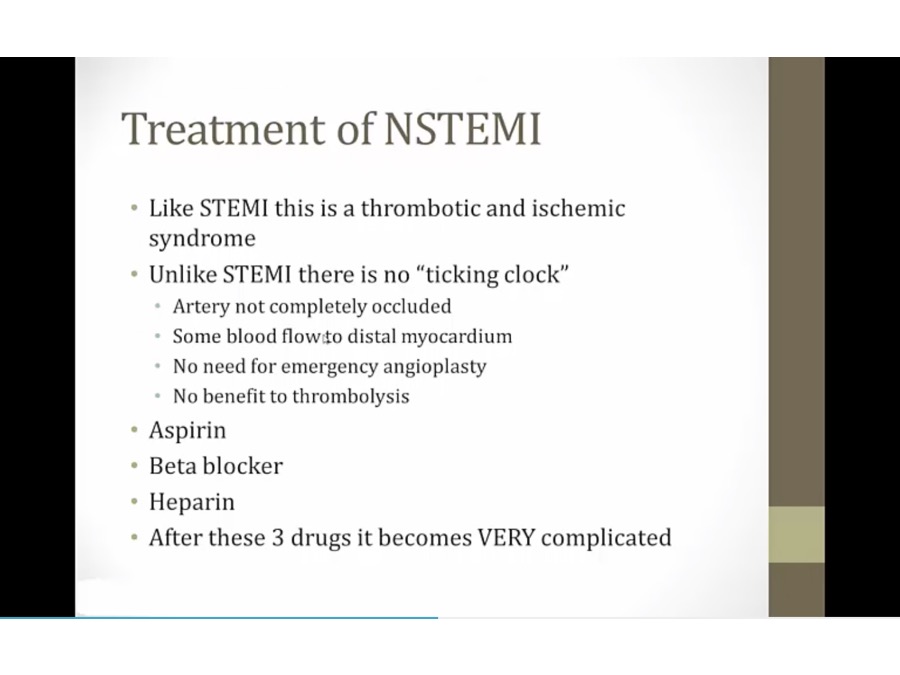
not important
Unstable Angina
_..
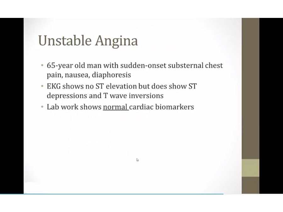
treated just like NSTEMI
Last updated