01 Cardiac Embryology
Heart Embryology
Heart contraction
Primitive heart tube
_Formed from Lateral folding directing the endocardial heart tubes to fuse., 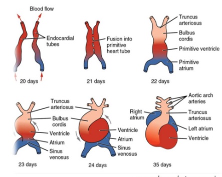
Cardiac looping
_ begins during the 4th week of gestation, and establishes a left-right polarity to the primitive heart tube. This process requires dynein., 

Heart beat
Primitive heart tube
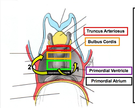
truncal-bulbar ridge top
left horn/left side: coronary sinus
right horn: RA 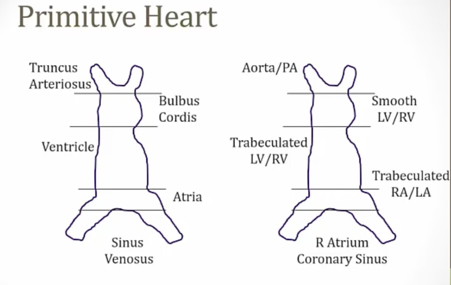
Truncus arteriosus
_Develops into the ascending aorta and pulmonary trunk.,
Bulbus cordis
_Develops into the conus arteriosus (smooth part of the right ventricle) and the aortic vestibule (smooth part of left ventricle)., 
Primitive ventricle
_Develops into trabeculated part of right and left ventricles.,
Primitve atrium
_Develops into the muscular (trabeculated) part of right and left atrium and the septum primum.,
Left horn of sinus venous
_Develops into the coronary sinus., (left side)
Right horn of sinus venous
_Develops into the smooth part of the right atrium., (right side)
Crista Terminalis
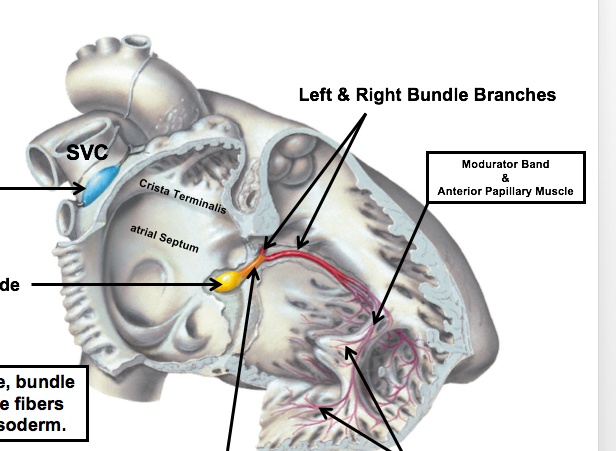
Superior Vena Cava
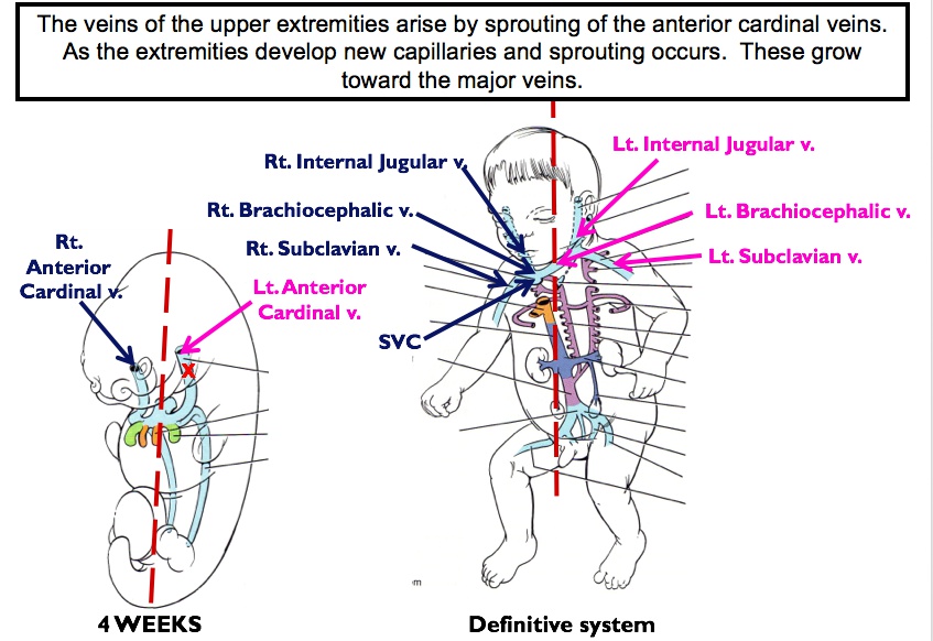
_Structures from outside the heart tube help form the adult heart structure, such as the right common cardinal vein and right anterior cardinal vein which develop into the superior vena cava., 
Primitive pulmonary vein
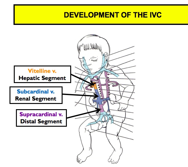
Inferior Vena Cava
Aorticopulmonary septum
_Neural crest cells migrate from the hindbrain to the primitive heart tube (Truncal bulbar ridge), which ultimately forms this., 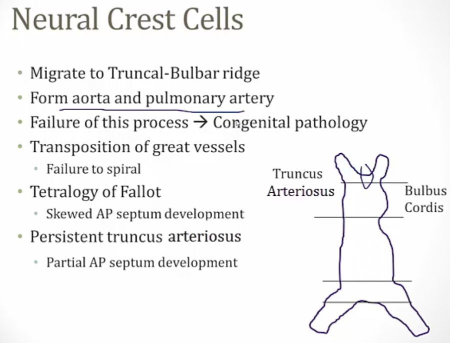
EDC of outflow tract
EDC of atrioventricular canal
Heart Septum Formation
Septum Primum
_Foramen primum is the first foramen. During the 4th gestational week, a septum separating the primitive atrium begins expanding from the superior interatrial space towards the endocardial cushions, forming septum primum.. 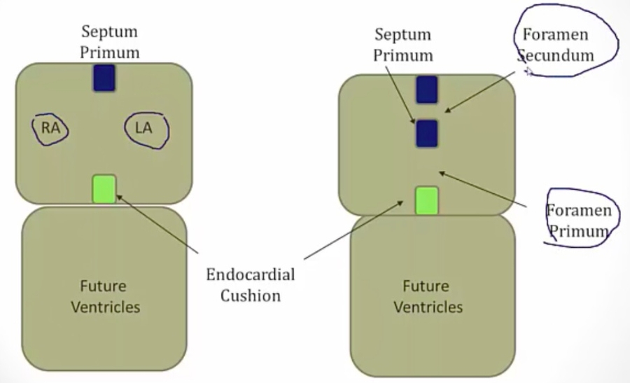
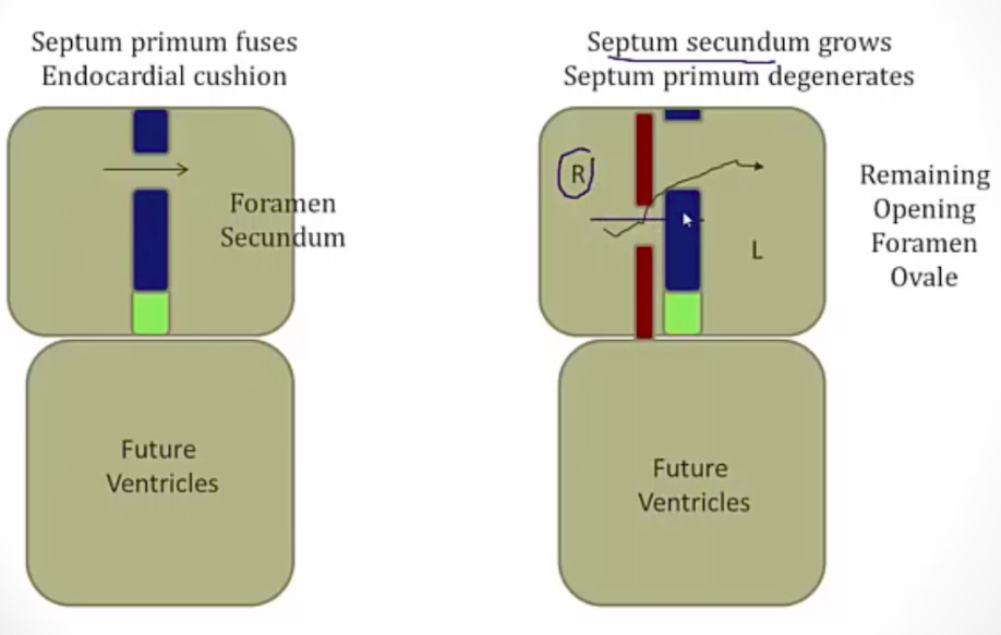
_The septum primum does not completely close against the endocardial cushions, leaving the initial opening, foramen primum. The septum primum then develops perforations that join to form foramen secundum. Septum foramen now has two holes in it..
Septum secundum and foramen ovale
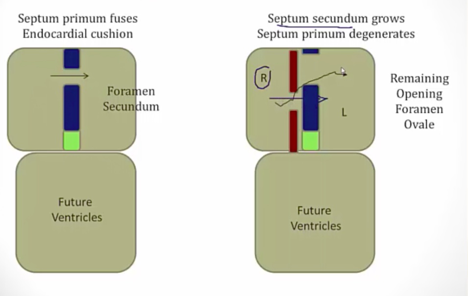
Foramen ovale
Fetal Circulation vs Changes in circulation at birth
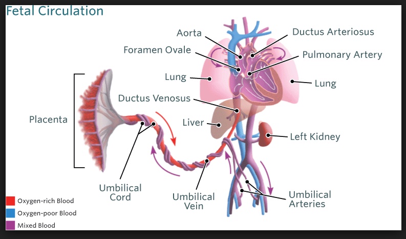
Ductus Arteriosus
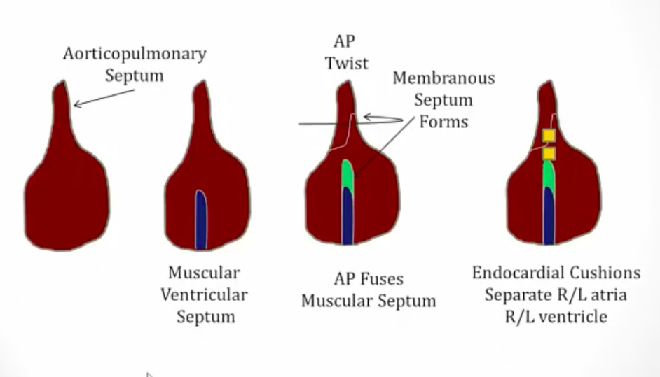
Ventricular septum development
Typically, the rotation of the aorticopulmonary septum and formation of the membranous interventricular septum closes the interventricular foramen.. 
Last updated