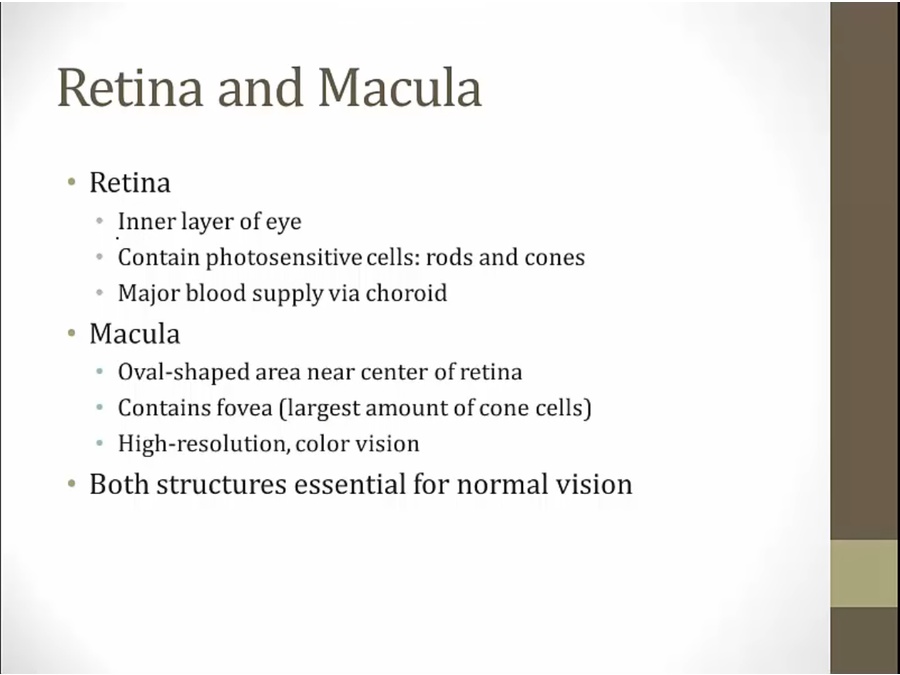
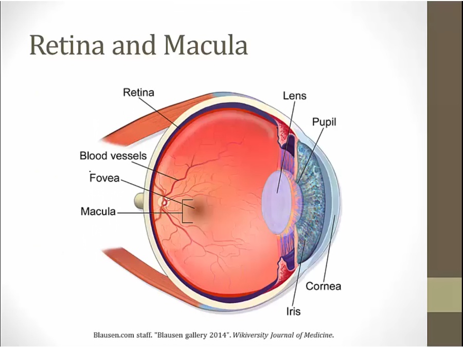
27 Retina
Structures
Pathology
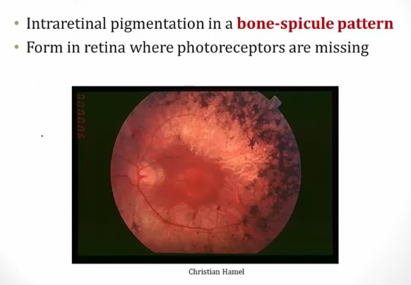
Retinitis Pigmentosa
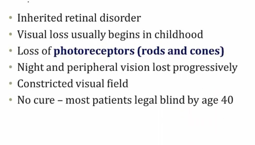
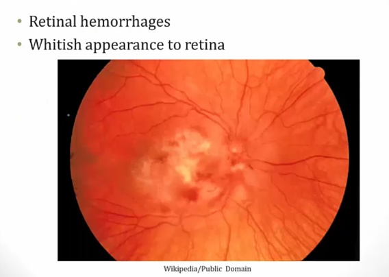
Retinitis
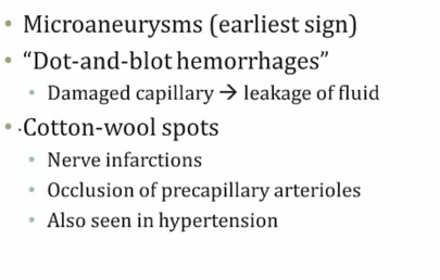
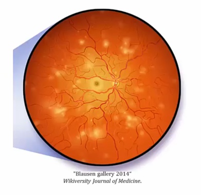
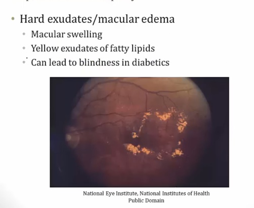
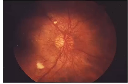
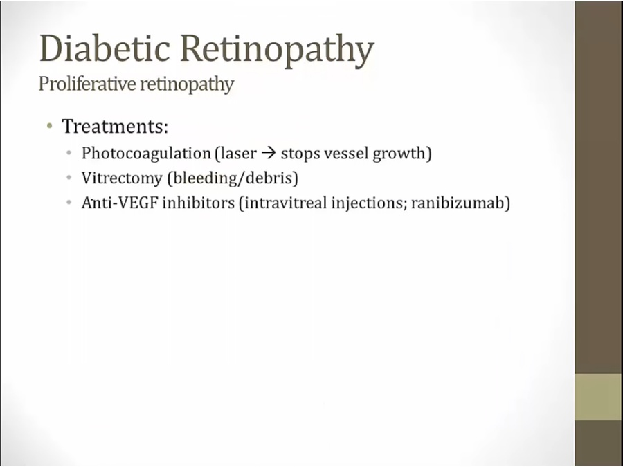
Diabetic Retinopathy
Nonproliferative
Proliferative
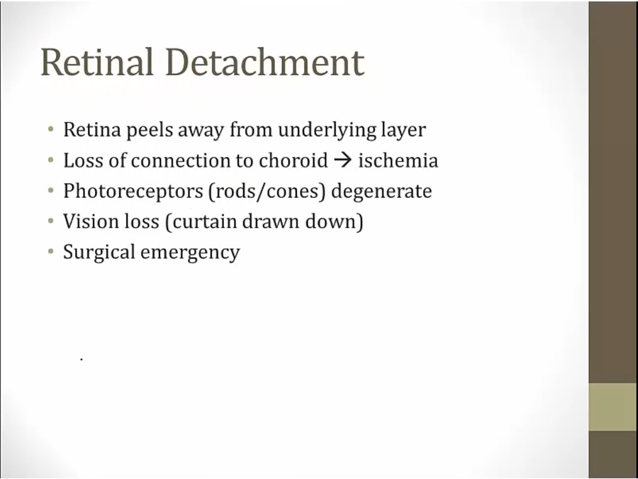
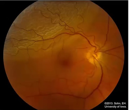
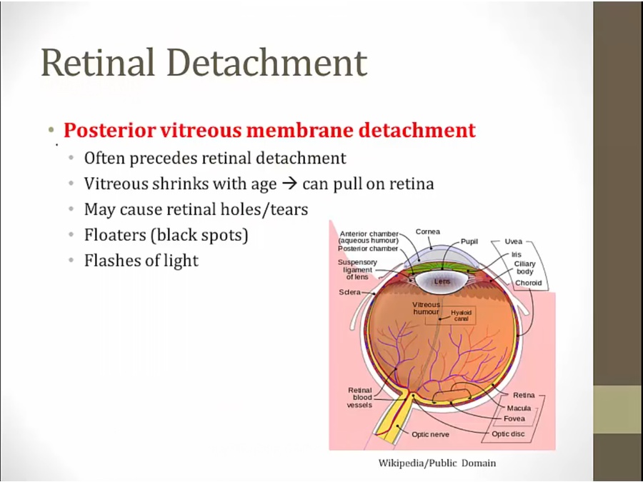
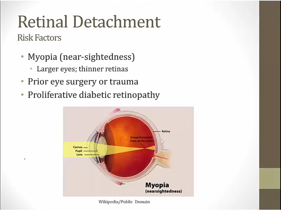
Retinal Detachment
Amaurosis Fugax
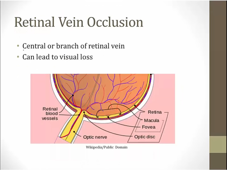
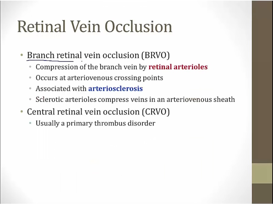
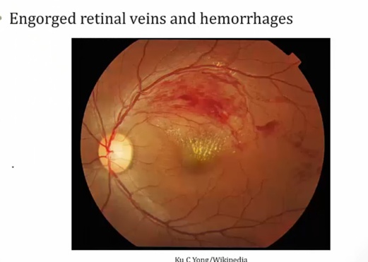
Retinal Vein Occlusion
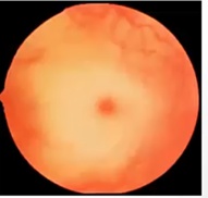
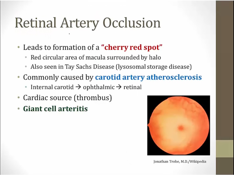
Retinal Artery Occlusion
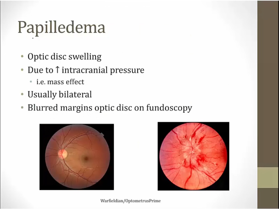
Papilledema
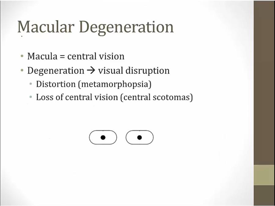
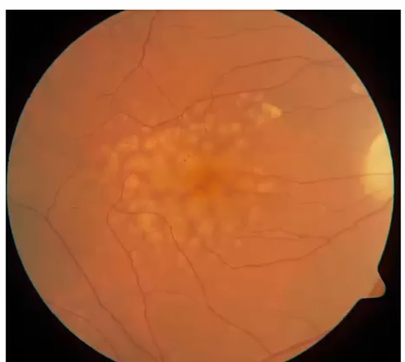
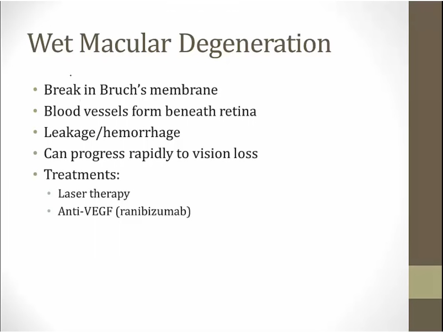
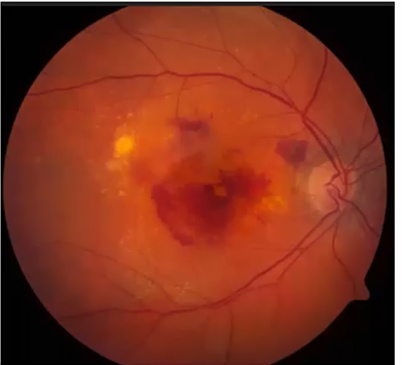
Macula Degeneration

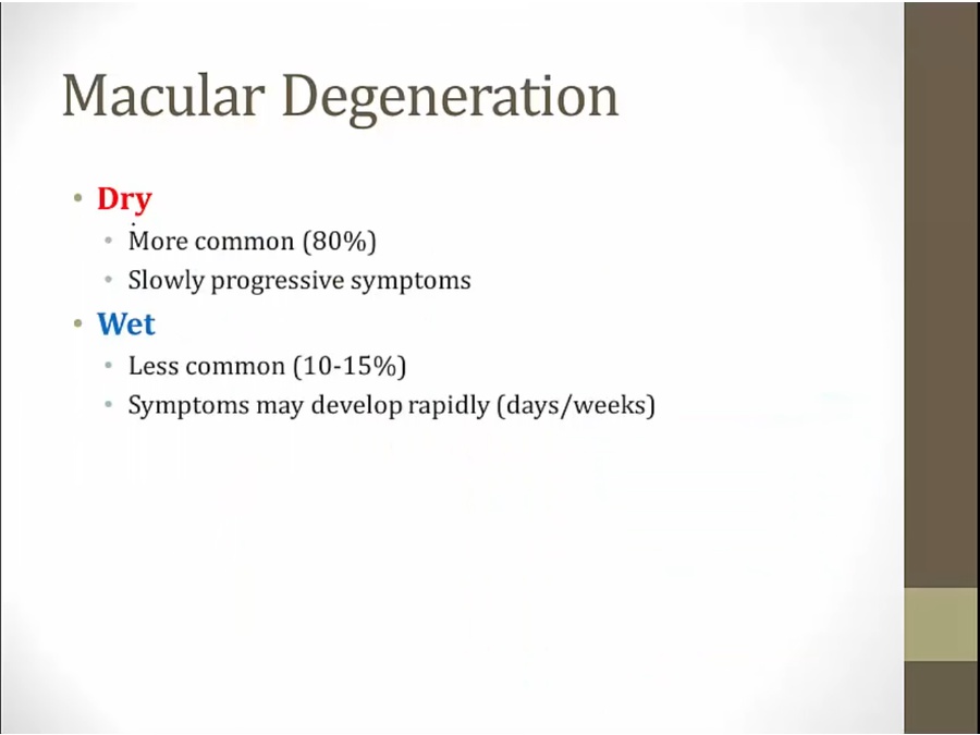
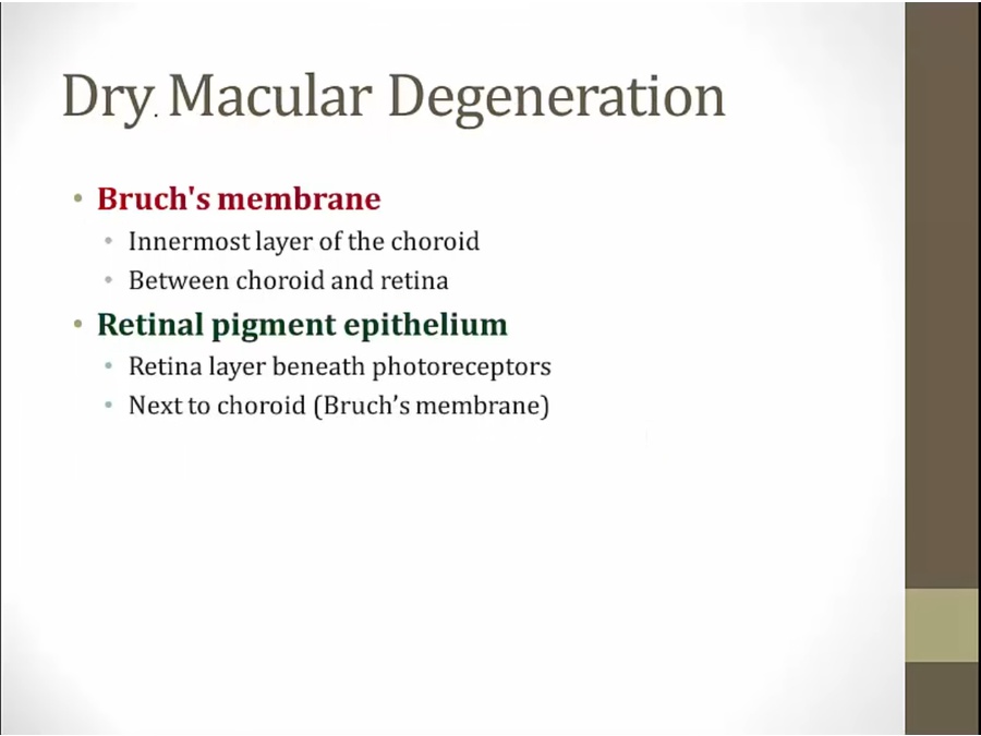
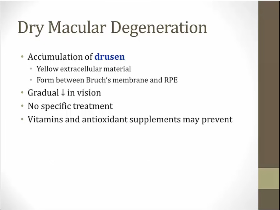
Dry

Wet
Last updated