20 PE
Pathogenesis
_..


femoral vein: thigh
popliteal vein: behind knee
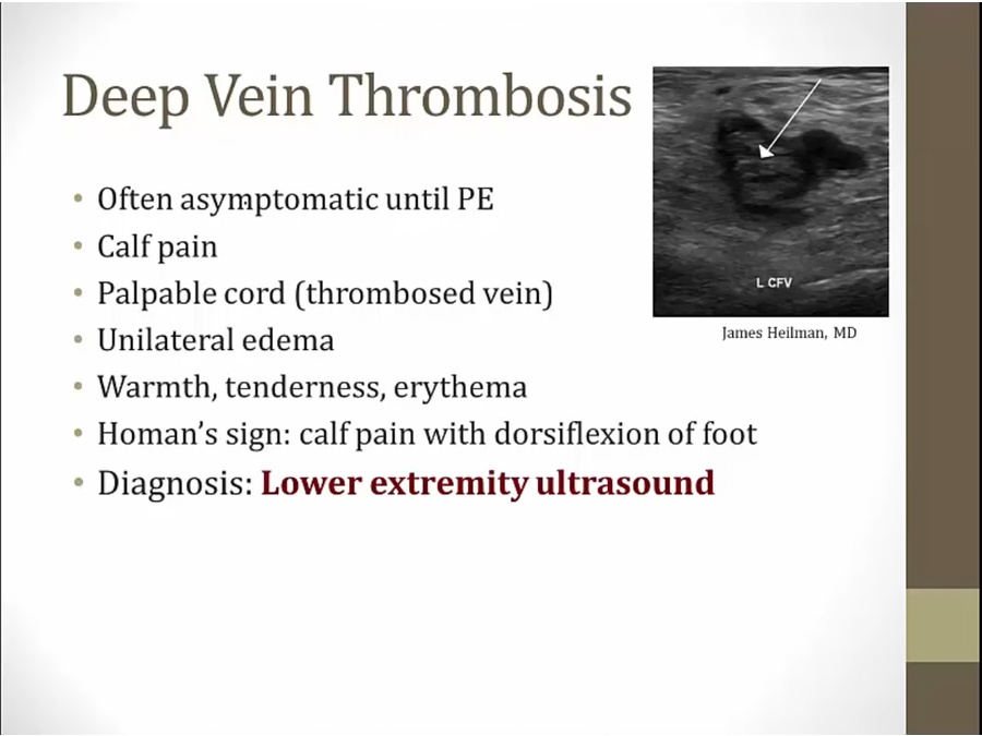
edema from poor venous outflow
Symptoms
_..
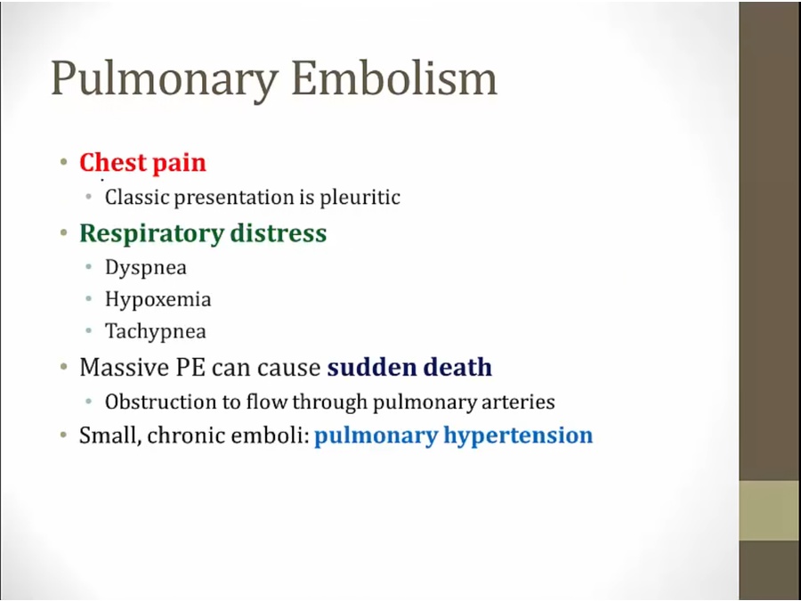
worse with deep breath
sudden death: so much obstruction of blood flow that dies from shock
Diagnosis
_..

CT contrast
thrombus in pulmonary artery

large PE crossing bifurcation
saddle emboli
pt likely to be in shock


classic EKG
deep S wave in lead 1, Q wave and inverted T wave in lead III
pseudo RBBB in V1, lower right: normal
sign of right heart strain: massive PE
in clinical practice and ER
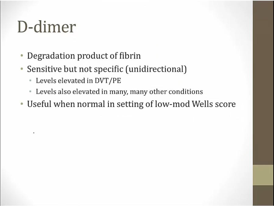
blood clots present and broken down by coagulation system: raise D-dimer
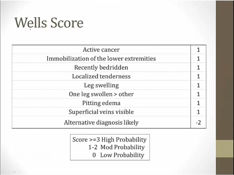
for ruling out DVT/PE with low Wells score

inhale nuclear tracer: left
inject nuclear tracer: right
see difference in V/Q
Treatment
_..

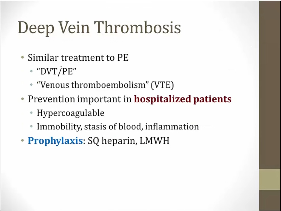
similar causes and treatment to PE
together aka VTE

Fat Embolism
_..

fat in bone marrow leak out
triad of symptoms in lung, CNS, skin

lung: respiratory failure
looks like ARDS
pt after trauma present with dyspnea, confusion, petechiae
Amnionic Fluid Embolism
_..


women shortly after delivering baby develops symptoms
biopsy shows fetal squamous and keratin debrid
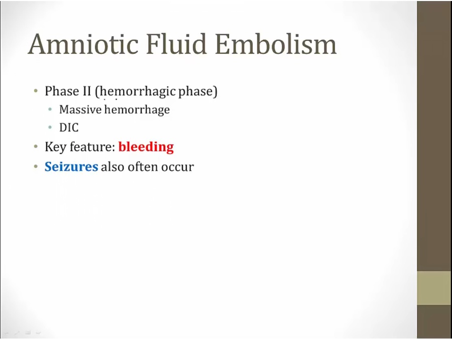
seizure can be seen in phase 1 or 2
Last updated