Breast
Embryogenesis
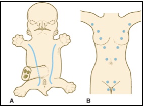
_Milkline/crest forms during 4th week. Milklines are formed from thickening of epidermis.. 
_Layers of epidermis:.. 
Germ layers
_Epidermis downgrowth into dermis. Ductal epithelial cells that make milk made from basalis layer of epidermis. Myoepithelial cells that contract to secrete milk made from mesenchymal (fibroblasts, mesoderm) around ducts..  Anything with milk production made by ectoderm
Anatomy
_Milk produced in the alveolus and stored in the lactiferous sinus. Contraction of myoepithelial cells forces milk out of the nipple. There are 9-10 lactiferous ducts per breast..


Glands of Montgomery
_Sebaceous glands of breasts. No hair follicles. Secretes lipoid substances that keep the breast moist., 
Blood Supply
_ Anterior intercostal.. 
Lymphatics
_.. 
Estrogen
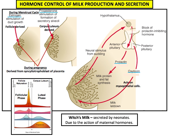
_stimulates lactiferous duct growth and breast tissue enlargement at puberty.,
Progesterone
_stimulates formation of secretory alveoli that produces the milk., 
_Estrogen and progesterone produced from follicle, corpus leuteum during ovulation, and from syncytiotrophoblast of placenta during pregnancy.. 
Lactogenesis
_Stages of milk production:
Lactogenesis, stage 1, late pregnancy: Alveolar cells differentiated from secretory cells and begin producing colostrom (fore milk), clear fluid of high protein/mineral concentration and low fat content
Stage 2, 2-3 days after birth: Colostrum switches to milk production (hind milk), higher fat content
Galactopoiesis, 9 days after: Milk production/secretion maintained by supply and demand
Involution, 40 days after: Milk production decrease as supplement added..

Prolactin
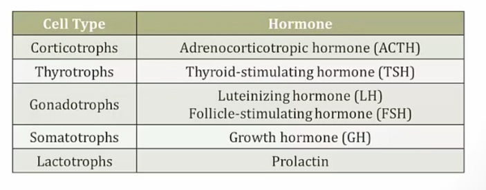
_Secreted by lactotrophs of the anterior pituitary and stimulates milk production as well as the development of milk-producing breast lobules.,

_ Aka luteotropin.,
_ Is a hormone named because of its role in lactation, and it is produced by:
Maternal adenohypophysis
Fetal adenohypophysis
Decidual tissue of the uterus..
_3 major actions:
Proliferation of mammary ducts at puberty and during pregnancy (with help of estrogen and progesterone).
Milk production and secretion in breast in response to suckling by inducing synthesis of milk components: lactose, casein (the protein of milk), and lipids.
Negative feedback on the hypothalamus causes ↓ GnRH (LH and FSH decrease) → inhibits ovulation. (Lactation amenorrhea).,
_During pregnancy the levels of prolactin, estrogen and progesterone increase. This increases prolactin secretion from lactotrophs. However, estrogen and progesterone also down-regulate prolactin receptors, preventing lactation from occurring during pregnancy. Also dopamine..
Inhibited by dopamine via binding to D2 receptors
Destruction of hypothalamus leads to high prolactin
Prolactin feedback on hypothalamus: increases dopamine release..
_At parturition, there is a precipitous drop in estrogen and progesterone, and at that point prolactin receptors are no longer down-regulated, allowing the high levels of prolactin seen during pregnancy to take action, thereby triggering lactation to occur..
_Secretory IgA antibody helps to reduce neonatal gut infections since neonates have underdeveloped gut immune system..
_Suckling is required to maintain milk production, because the increased nerve stimulation increases oxytocin and prolactin levels..
_The suckling infant stimulates hypothalamus to produce oxytocin that increases milk secretion. This is called letdown reflex..
_Reflex arc activates 3 areas:
inhibit dopamine (prolactin increase)
increase oxytocin secretion (contraction of breast myoepithelial cells)
uterine contraction (pain)..
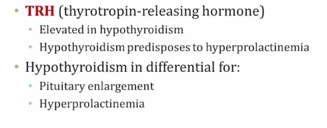
_Prolactin is increased by TRH:..

Oxytocin
_A nonapeptide that is primarily produced by the paraventricular nuclei within the hypothalamus, and secreted from the posterior pituitary. Like ADH, oxytocin travels to the posterior pituitary via carrier proteins called neurophysins.,
_Antidiuretic hormone is homologous to oxytocin. These nonapeptides only differ in structure by two amino acids..
_The major stimulus for secretion is suckling. Other positive stimuli include dilation of the cervix and orgasm.,

_The primary effects are milk ejection and uterine contraction. Causes milk ejection by inducing contraction of myoepithelial cells in the breast.,

_Clinical correlation: Oxytocin (Pitocin) can be given to reduce postpartum bleeding because it causes uterine contraction..
_Can have several adverse effects. These include:
Hyponatremia and seizures (due to anti-diuretic properties similar to antidiuretic hormone)
Subarachnoid hemorrhage
Uterine rupture in pregnant patients.,
_..
From
Effect
Prolactin
Anterior pituitary
milk synthesis
Oxytocin
Posterior pituitary
milk contraction by myoepithelial cells

Last updated