Derm
Cutaneous Fungla Infections
Cutaneous fungal infections are typically characteristic for specific areas of the body and are frequently associated with:
Warm or moist environments
Diabetes mellitus
Obesity
Recent antibiotic use
Tinea
Tinea versicolor, also known as pityriasis versicolor, is a cutaneous fungal infection caused by yeasts in the genus Malassezia (formerly known as Pityrosporum). It is most commonly caused by Malassezia globosa.
Malessezia, the causative genus of tinea versicolor, is a dimorphic fungus that is part of the normal skin flora and is not contagious.
Pathologic tinea versicolor is most common in the summer due to hot, humid weather.
Additional risk factors include the use of topical skin oils and hyperhidrosis (excessive sweating).
Patients with tinea versicolor classically present with hypopigmented lesions that fail to tan despite summer sun exposure.
Tinea versicolor presents with hypopigmented, hyperpigmented, or salmon-colored macules on the trunk and proximal upper extremities from the degradation of lipids that produces acids that damage melanocytes.


Diagnosis of tinea versicolor is made with a potassium hydroxide (KOH) preparation. Both hyphae and yeast cells are evident in a pattern that is described as “spaghetti and meatballs.”
First-line treatments for tinea versicolor include:
Selenium sulfide
Zinc pyrithione
Topical antifungals (e.g. ketoconazole 2% cream)
Dermatophyte
Dermatophyte (non-Malassezia tinea) infections, also known as ringworm, are cutaneous fungal infections caused by:
Epidermophyton
Trichophyton
Microsporum
Lesions are pruritic, erythematous, scaly plaques that have a characteristic central clearing. This gives the lesion the appearance that a ring surrounds it.
Dermatophyte infections are named based on the location of the lesion as follows:
Head, scalp: tinea capitis
Torso: tinea corporis
Groin: tinea cruris
Feet: tinea pedis
Nail bed: tinea unguium (onychomycosis)

The diagnosis can be made with a potassium hydroxide preparation, which shows hyphae.
Treatment consists of:
Topical antifungal medication for multiple weeks with oral antifungal in resistant cases
Oral griseofulvin or terbinafine for tinea capitis
Oral terbinafine for onychomycosis
Intertrigo
Intertrigo is as an inflammatory condition of skin folds (intertriginous areas). It is most commonly caused by Candida albicans.

Lesions are pruritic, painful, erythematous plaques most commonly found in skin creases.
The diagnosis can be made with a potassium hydroxide preparation, which shows budding yeasts with septate hyphae and pseudohyphae.
Treatment consists of topical antifungal medication and topical corticosteroids.
Atopic Dermatitis
Atopic dermatitis, otherwise known as eczema is a chronic inflammatory skin disease of unknown etiology that results in pruritus, scaling, crusting, and lichenification of the skin.
Eczema primarily occurs before the age of 5 years old and is commonly associated with other atopic disorders such as asthma, allergic rhinitis, and food allergies. Persistent eczema in the pediatric population may warrant allergy testing.
Patients with eczema classically present with relapsing and remitting red, scaly, crusting lesions on the cheeks, face, flexural creases, or extensor surfaces.
Eczema is diagnosed primarily on history and presentation alone. There are no specific diagnostic modalities for eczema.
On physical examination patients will typically present with erythematous, dry, scaly, crusted lesions affecting the face and flexural creases. Lichenification typically occurs in early childhood.
Eczema may result in:
Scarring
Lichenification
Secondary bacterial infections from excessive scratching
Eczema herpeticum due to HSV superinfection
Eczema herpeticum may affect multiple organs and may appear as umbilicated vesicles on top of healing atopic dermatitis. It is treated with acyclovir/valacyclovir or foscarnet in cases of acyclovir resistant HSV.
Topical steroids and emollients such as petrolatumare currently the mainstay treatment in eczema. Keeping the skin moisturized helps prevent excessive dryness and pruritus.
Tacrolimus and Pimecrolimus are immunomodulators used in a topical formulation for the treatment of moderate to severe eczema.
Bullous Pemphigoid
Bullous pemphigoid is an autoimmune subepidermal blistering disease. Bullae form between the epidermis and the dermis. The pathology is not completely understood, but thought to be due to autoantibodies damaging subepidermal connections within the skin.
Bullous pemphigoid is a rare disorder that primarily occurs in individuals >60 years old, with equal incidence in men and women.
Some medications such as furosemide, NSAIDs, ACE inhibitors, and antibiotics have been associated with the development of bullous pemphigoid.
Patients with bullous pemphigoid classically present with a prodromal period, which includes severe pruritus with plaque-like lesions. Following the prodromal period, which can lasts for weeks, tense fluid-filled blisters begin to appear.
The most worrisome complication in bullous pemphigoid is secondary bacterial infection, especially in patients who are bedbound. Other complications include fluid loss and decreased activities of daily livingwhen the palms and soles are affected.
The first-line treatment for bullous pemphigoid is a high-potency topical corticosteroid such as clobetasol.
When topical steroids are not able to control the disease, oral corticosteroids may be used.
Those patients who are refractory to medical therapy, Intravenous Immunoglobulin (IVIG)is utilized in the treatment of bullous pemphigoid.
Patients are advised to not pop their blisters in order to reduce the risk of infection.
Skin biopsy with Direct Immunofluorescence (DIF) is the diagnostic modality of choice in bullous pemphigoid. DIF detects IgG and Complement C3 within the subepidermal junction.
On physical exam patients will present with tense fluid filled blisters seen more on the flexor surfaces. Nikolsky’s sign (rubbing of skin resulting in exfoliation of outermost layer) will be negative.
Oral lesions are very rare with bullous pemphigoid (more typically seen with pemphigus vulgaris).
Pemphigus Vulgaris
Pemphigus vulgaris is an autoimmune, intraepithelial blistering disorder that affects the skin and mucus membranes. Although the actual cause of pemphigus vulgaris is unknown, it is thought to be the result of antibodies attacking the attachments within the epidermis.
Pemiphigus vulgaris differs from bullous pemphigoid in that it most often always has mucosal involvement, has flaccid blisters, and involves the intraepithelial layer.
Pemphigus vulgaris is an extremely rare disorder that has an average age of onset between 40 to 60 years old, and an equal incidence between men and women.
Complications associated with pemphigus vulgaris include secondary bacterial infection, malnutrition, dehydration, and scarring.
The drug of choice in treating pemphigus vulgaris is systemic corticosteroids. Other adjuvant therapies include azathioprine and mycophenolate mofetil.
Almost all patients present with mucosal involvement, which typically includes painful blistering and ulcerations of the lips and oral mucosa. Blisters on the skin tend to rupture very easily, so some patients will present with bright red skin lesions.
Mucosal lesions tend to occur first, with the cutaneous lesions occurring weeks to months after.
The gold standard in diagnosing pemphigus vulgaris is a skin biopsy with Direct Immunofluorescence (DIF), which will reveal IgG and Complement C3 deposition within the epidermis.
On physical exam the patient’s oral mucosa will be extremely tender to the touch and typically have several erosions on their lips, gums, and cheek lining.
Their blisters are very fragile and tend to burst with light touch (positiveNikolsky sign), which will leave a painful erythematous lesion behind.
Contact Dermatitis
Contact dermatitis is a localized inflammatory response to a variety of chemical and physical irritants, which results in edema, erythema, and damage to the skin. It is further divided into irritant contact dermatitis and allergic contact dermatitis.
Allergic contact dermatitis is a type IV hypersensitivity reaction. Irritant contact dermatitis is mediated by proinflammatory cytokines released due to harmful physical or chemical stimuli.
The actual level of irritation depends on the amount/length of exposure, chemical properties, and environmental exposure. Chemical irritants include detergents, cleaning agents, alcohol based hand sanitizer, soap, bleach, benzoyl peroxide, and excessive water exposure). Physical irritants include fiberglass, plant parts (e.g. thorns, serrated leaves), soil, and metal/wood shavings.
Work environment plays a significant role in patients presenting with contact dermatitis. For instance, custodial positions, health-care workers, as well as metal and woodshop positions all experience repeated chemical and physical exposures.
The clinical manifestations of contact dermatitis include dry, pruritic, red irritated skin and vesicles. Calluses, skin atrophy and even skin necrosis may develop from harsher irritants.
Contact dermatitis is primarily diagnosed by physical exam and history of irritant exposure. A skin biopsy and allergy patch testing can be used to help rule out allergic contact dermatitis and other disorders such as psoriasis.
On physical examination, a patient’s skin can show:
Dryness
Pruritic vesicles
Erythema
Fissuring
Blisters
Atrophy and even necrosis
Contact dermatitis may result in scarring, loss of functionality, de-sensitization, secondary bacterial infection, and even skin loss.
Treatment of contact dermatitis includes the identification and avoidance of irritants. Treatment also includes emollients, barrier creams, appropriate glove use, and in severe cases topical corticosteroids.
Emollients help reduce skin irritation and provide a barrier to further irritation.
Topical steroids are used in more severe cases in which there is prominent skin thickening.
Erythema Multiforme
Erythema Multiforme (EM) is an acute cutaneous type IV hypersensitivity condition that is immune-mediated and characterized by distinctive target-like lesions.
The two most important infectious causes of erythema multiforme are HSV (most common overall) and Mycoplasma pneumoniae.
Medications of note that have been associated with EM include:
Penicillins
Sulfonamides
NSAIDs
Oral contraceptives
Anticonvulsants
The condition affects males more than females and typically occurs in people 20-40 years of age.
The target lesions seen in erythema multiforme appear as an erythematous center surrounded by a pale inner ring followed by an erythematous outer ring.
EM may also present with:
Malaise
Myalgias
Macules
Plaques or vesicles on extremities (notably palms or soles)
Skin biopsy findings associated with erythema multiforme include increased lymphocytes and necrotic keratinocytes. Lab work will show increased eosinophils.
Erythema multiforme can be self limiting, but treatment strategies include:
Stopping the offending agent
Corticosteroids
Analgesics
(If present) treat HSV or mycoplasma
Erythema Nodosum
Erythema Nodosum is an inflammation of subcutaneous fat that results in painful erythematous nodules, usually in a pretibial location.
The inflammation is thought to be a result of a delayed hypersensitivity reaction to antigens associated with various infectious agents, drugs, or other diseases.
Erythema nodosum is most common amongst women ages 15-40.
The single most frequent condition associated with the development of erythema nodosum is Streptococcal pharyngitis.
The typical presentation of erythema nodosum involves painful erythematous nodules, usually on the anterior portion of the tibia with possible malaise, fatigue, and polyarthralgia.
The diagnosis is usually made clinically, but a biopsy can be performed to confirm erythema nodosum in atypical cases, and will show fatty inflammation.
Other laboratory findings include a positive antistreptolysin O antibody (if associated with a strep infection) and an increased erythrocyte sedimentation rate.
In most cases, erythema nodosum will either resolve on its own or resolve following treatment of the underlying disorder.
Pharmacologic management of erythema nodosum may consist of:
NSAIDs
Potassium iodide
Corticosteroids
Molluscum
Molluscum contagiosum is a poxvirus that causes a viral skin infection resulting in a chronic localized infection.
The virus spreads person to person by direct contact and can even cause self-infection if lesions are scratched or touched. It can also spread via fomites.
The infection is most commonly seen in children and patients that are HIV positive.
The skin infection caused by molluscum contagiosum is characterized by painless, shiny papules with a central umbilication that are usually less than 5 mm in diameter.
The most common locations the infection is found in children include:
Face
Back
Chest
Extremities
In adults, the infection is most often found in the perineal region.
Molluscum dermatitis is common, and consists of eczematous patches and plaques that surround the molluscum contagiosum lesions.
The diagnosis of molluscum contagiosum is usually made clinically based on the appearance of the lesions.
If necessary, histological specimens can be viewed with Giemsa or Wright stains and will show large inclusion bodies.
Molluscum contagiosum is most often self-limited, but treatment options include:
Cryotherapy
Curettage
Cantharidin, which is a topical blistering agent
Topical trichloroacetic acid, used as a chemical peel
Pityriasis Rosea
Pityriasis rosea is an acute, mild inflammatory skin disease that is characterized by papular lesions on the trunk and proximal areas of the extremities and is thought to arise from a viral origin.
It occurs more often in older children and young adults and more frequently in women than men.
Pityriasis rosea classically begins with a 2-5 cm "herald" patch: a round salmon-colored lesion on the trunk that scales and clears centrally, leaving a "collarette" of scale. Days to weeks later, smaller lesions with similar characteristics appear on the trunk and extremities.
Pityriasis rosea lesions classically have a "Christmas tree" distribution on the trunk because the long axes of the oval plaques are oriented along the lines of skin cleavage.
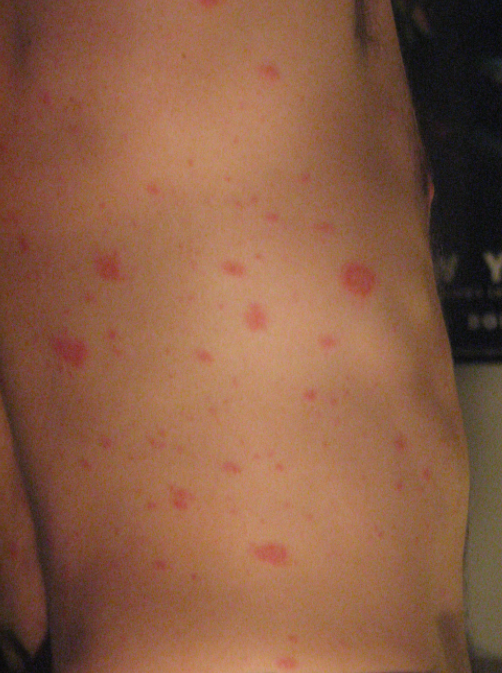
Pityriasis rosea is usually asymptomatic with the exception of local pruritus, which can be treated with medium-potency topical corticosteroids.
Pityriasis rosea is usually diagnosed clinically, but sometimes a potassium hydroxide (KOH) examinationof the herald patch scale is performed to rule out tinea corporis (both lesions can appear as a plaque with central clearing and peripheral scale).
KOH examination of tinea corporis will show dermatophyte hyphae, while these will be absent in pityriasis rosea.
The papules and plaques typically resolve in four to six weeks.
Cutaneous manifestations associated with PCT include:
Chronic blistering skin lesions on sun exposed areas of the body (most commonly on the back of the hands) because the porphyrins that accumulate are photosensitizing
Hyperpigmented skin
Blisters, bullae, and increased fragility
Hirsutism (especially on cheeks and forearms)
Hepatic manifestations associated with PCT include:
Mild elevations in liver enzymes (ALT and AST)
Advanced liver disease in older patients with recurrent disease
Increased risk of cirrhosis and hepatocellular carcinoma
Although the characteristic blistering skin lesions are highly suggestive of porphyria cutanea tarda, the diagnosis should be supported biochemically by demonstrating:
Increased total plasma porphyrin
Increased urine porphyrin
Decreased uroporphyrinogen decarboxylase
Elevated AST and ALT
The preferred treatment of PCT is repeated phlebotomy (effective in nearly all patients).
Other treatments that can be used include:
Low dose chloroquine or hydroxychloroquine (for patients who do not tolerate phlebotomies)
Sunscreen use (to mitigate photosensitivity)
Avoidance of triggers (alcohol, tobacco, estrogens, iron supplements)
Porphyria Cutanea Tarda
Porphyria cutanea tarda (PCT) is a deficiency of hepatic uroporphyrinogen decarboxylase(UROD) that results in chronic blistering skin and liver manifestations.
The deficiency isacquired in 80% of cases and autosomal dominant in 20% of cases.
Risk factors associated with PCT include the use of hepatotoxic agents, such as:
HCV infection
Alcohol
Smoking
Estrogen (e.g. OCPs)
Iron overload (e.g. hereditary hemochromatosis)
Notable pharmacology correlate: Barbiturates (or other P450 inducers) result in the consumption of heme, which leads to a loss of heme’s negative feedback on ALA synthase (the rate limiting step of heme biosynthesis), which causes an accumulation of heme intermediates. Accumulation of these intermediates causes acute exacerbations of porphyrias.
Warts
Warts (verrucae) are benign epithelial tumors caused by local infection by human papillomavirus (HPV).
HPV 1 is typically associated with plantar warts and HPV 6 and 11 are typically associated with anogenital warts.
HPV warts are spread by direct skin-to-skin contact.
Patients with warts will have well defined lesions consisting of thickened epithelium that can either be flat or raised and are occasionally tender to palpation.
The diagnosis is made based on clinical appearance, but scrapings of hyperkeratotic debris will show thrombosed capillaries often referred to as“seeds.”
Warts are self-limiting for many patients, but treatment in persistent cases can include:
Salicylic acid (safest and most efficacious)
Liquid nitrogen (cryotherapy)
Trichloroacetic acid
Curettage and desiccation
Laser removal
The Gardasil vaccine (tetravalent against HPV types 6, 11, 16, 18) is now FDA approved for both males and females.
Complications associated with warts are based on the specific subtype of HPV, such as type 16 and 18 which are associated with cervical cancer.
Last updated